
|
Fig. S3
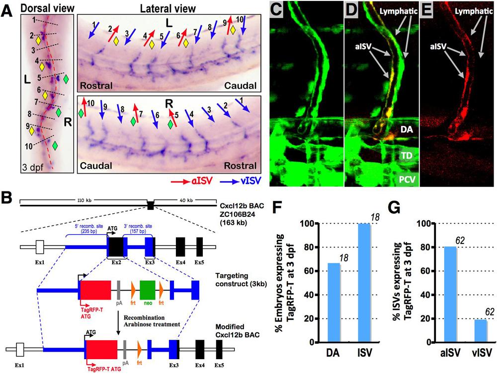
(related to Figure 3). Examining cxcl12b expression using whole-mount in situ hybridization and recombineered Cxcr12b BAC constructs.
(A) Determination of the arterial-venous identity and cxcl12b expression of each trunk ISV. An example of how the presence (or absence) of cxcl12b expression in each individual intersegmental blood vessel (ISV) is assessed in an animal in which the arterial or venous identity of the same ISV has previously been determined. Transmitted light imaging is used to determine the direction of flow in each ISV, on both sides of the trunk in a 3 dpf zebrafish larvae (Movie S2; focusing up and down to visualize the vessels on either side of the trunk). Dorsal-ward flow indicates arterial (aISV) identity, ventral-wards flow indicates (vISV) venous identity. After recording the arterial or venous identity of each of the first 10 ISV on either side of the trunk, animals are fixed and each animal is separately subjected to whole-mount in situ hybridization (WISH) probing for cxcl12b. The presence or absence of cxcl12b expression in each of the ISV is then assessed blindly, examining from a dorsal (left), left lateral (top right), and right lateral (bottom right) perspective. The cxcl12b-positive ISVs are noted with yellow (left side) or green (right side) diamonds. Following determination of cxcl12b expression status, comparison is made to which vessels had previously been recorded to be aISV (red arrows in right panels) or vISV (blue arrows in right panels). The expression of cxcl12b strongly correlates with aISV rather than vISV identity. Red dashed line in dorsal view denotes the midline. (B) Construction of a targeted BAC construct driving TagRFP-T expression from cxcl12b promoter-enhancer sequences. (C-E) Confocal images of a 3.5 dpf Tg(fli1a:EGFP)y1 (green) larva injected with a recombineered BAC clone with TagRFP-T (red) driven by the cxcl12b promoter. TagRFP-T is detected in aISV, DA, but not in ISLV. (C) Green channel (EGFP) only. (D) Merged image. (E) Red channel (TagRFP-T) only. (F) Quantification of TagRFP-T expressing cell types observed in recombineered Cxcl12b BAC-injected Tg(fli1a:EGFP)y1 embryos at 3 dpf. Numbers above the bars show the number of TagRFP-T expressing embryos counted. (G) Quantification of TagRFP-T-expressing aISVs or vISV in embryos that have TagRFP-T-positive ISVs in F. Numbers above the bars show the number of TagRFP-T expressing ISVs counted.
Reprinted from Developmental Cell, 22(4), Cha, Y.R., Fujita, M., Butler, M., Isogai, S., Kochhan, E., Siekmann, A.F., and Weinstein, B.M., Chemokine Signaling Directs Trunk Lymphatic Network Formation along the Preexisting Blood Vasculature, 824-836, Copyright (2012) with permission from Elsevier. Full text @ Dev. Cell