
|
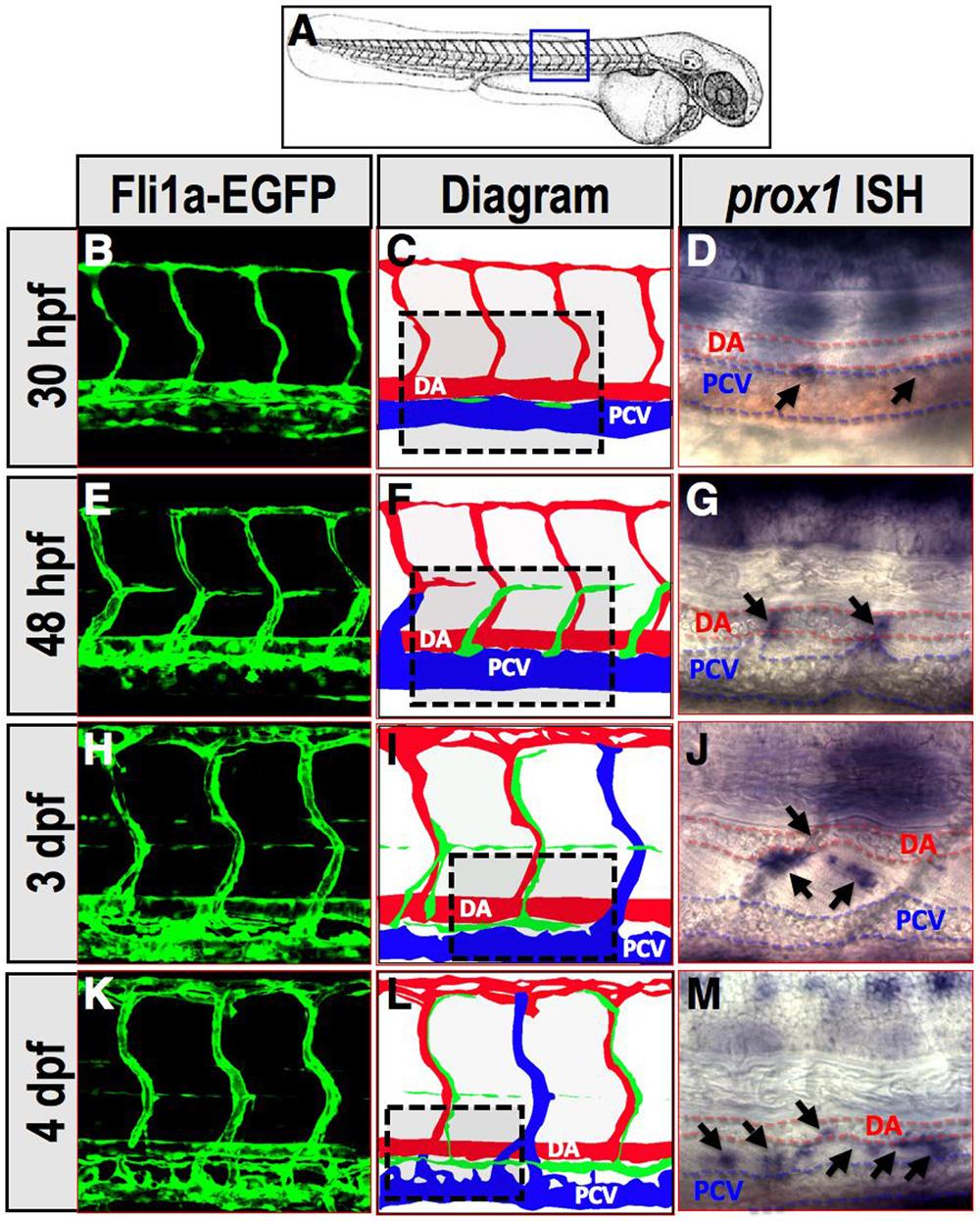
Fig. S1
(related to Figure 1). prox1 expression marks lymphatic progenitors and lymphatic endothelial cells throughout trunk lymphatic network development
(A) Diagram of a zebrafish embryo with a blue box highlighting the region shown in B-M. (B, E, H, and K) Confocal images of trunk vessels in Tg(fli1a:EGFP)y1 embryos and larvae at the stages indicated at left. (C, F, I, and L) Illustrative diagrams of the trunk vessels visualized in each of the confocal images at left. Red, arterial blood vessels (or primary intersegmental blood vessel (ISV) whose identity if as yet undetermined, in C); blue, venous blood vessels; green, prox1-positive lymphatic progenitors and developing lymphatic vessel (LV) segments. Box shows approximate area of whole-mount in situ hybridization (WISH) images at right. (D, G, J, and M) Transmitted light images of zebrafish embryos and larvae (at the stages noted at left) subjected to WISH, probing for prox1. Expression of prox1 is detected in both developing LV segments (G, J, and M) as well as in selected cells within the posterior cardinal vein (PCV) (marked with dab2-fluorescin probe, red staining) prior to emergence of lymphatic progenitors from these blood vessels (D).
Reprinted from Developmental Cell, 22(4), Cha, Y.R., Fujita, M., Butler, M., Isogai, S., Kochhan, E., Siekmann, A.F., and Weinstein, B.M., Chemokine Signaling Directs Trunk Lymphatic Network Formation along the Preexisting Blood Vasculature, 824-836, Copyright (2012) with permission from Elsevier. Full text @ Dev. Cell