
|
Fig. 4
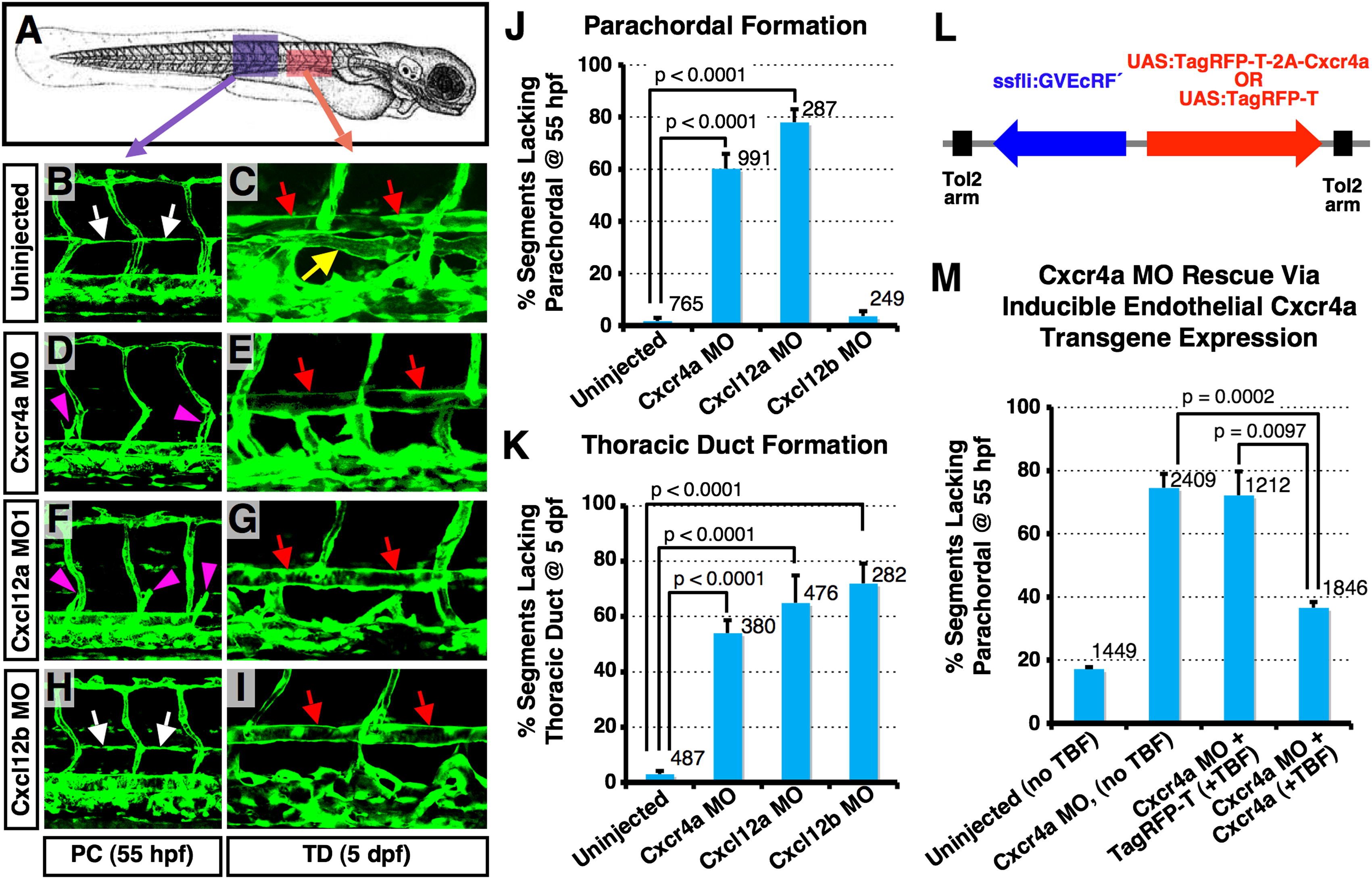
Lymphatic Network Formation Is Disrupted in Chemokine Morphants (A) Diagram of a zebrafish embryo (a purple box, the region shown in B, D, F, and H; a red box, the region shown in C, E, G, and I). (B–I) Confocal images of trunk vessels in 55 hpf (B), (D), (F), and (H) or 5 dpf (C), (E), (G), and (I) Tg(fli-EGFP)y1 embryos that were either not injected, or injected with MOs indicated at left. (J) Quantification of the PC formation defect in 55 hpf MO-injected embryos (as shown in B, D, F, and H). Values are the mean ± SEM. Numbers of counted PC segments are indicated above the bars. (K) Quantification of the TD formation defect in 5 dpf MO-injected embryos (as shown in C, E, G, and I). Values are the mean ± SEM. Numbers of counted TD segments are indicated above the bars. (L) Diagram of Cxcr4a rescue constructs for spatiotemporal overexpression of either TagRFP-T + Cxcr4a or TagRFP-T alone (arrows, direction of gene expression). (M) Quantification of the PC formation defect in control (column 1) or Cxcr4a morphant animals (columns 2–4), some of which were coinjected with transgenes for ecdysone-inducible endothelial-specific overexpression of either TagRFP-T (column 3) or Cxcr4a (column 4). Values are the mean ± SEM. Numbers of counted PC segments are indicated above the bars. White arrows, PC; yellow arrow, TD; red arrows, DA; magenta arrowheads, secondary sprouts migrating to the level of the HM. See also Figure S4.
Reprinted from Developmental Cell, 22(4), Cha, Y.R., Fujita, M., Butler, M., Isogai, S., Kochhan, E., Siekmann, A.F., and Weinstein, B.M., Chemokine Signaling Directs Trunk Lymphatic Network Formation along the Preexisting Blood Vasculature, 824-836, Copyright (2012) with permission from Elsevier. Full text @ Dev. Cell