
|
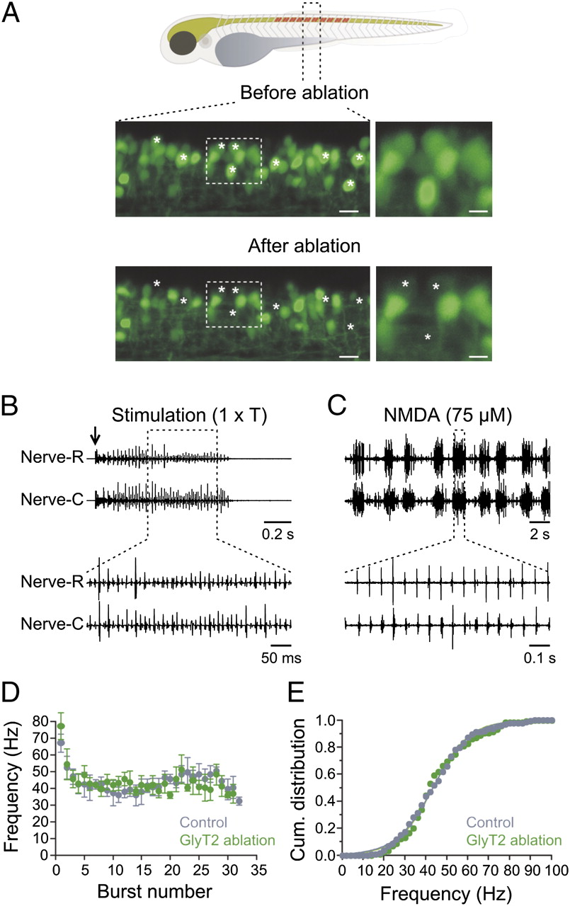
Fig. 4
Ablation of glycinergic (GlyT2) interneurons does not alter swimming activity. (A, Upper) Drawing of a zebrafish showing the region where GlyT2 interneurons are ablated. (Lower) Confocal reconstruction of 1.5 segment of the spinal cord before and after photoablation of GlyT2 interneurons. Asterisks indicate the position of the ablated interneurons. (Right) High magnification of the regions indicated by the dashed boxes. (Left scale bar, 10 μm and Right scale bar, 5 μm.) (B) Electrical stimulation at threshold intensity (1 × T; arrow) induces a bout of swimming activity in animals with GlyT2 interneurons ablated. (C) Application of a lower NMDA concentration (75 μM) induces swimming activity both in the region of the ablation and in more caudal segments. (D) Burst frequency and duration of the swimming bout recorded in animals with GlyT2 interneurons ablated is similar to control animals. (E) Cumulative distribution of the burst frequency in control and in animals with GlyT2 interneurons ablated.