
|
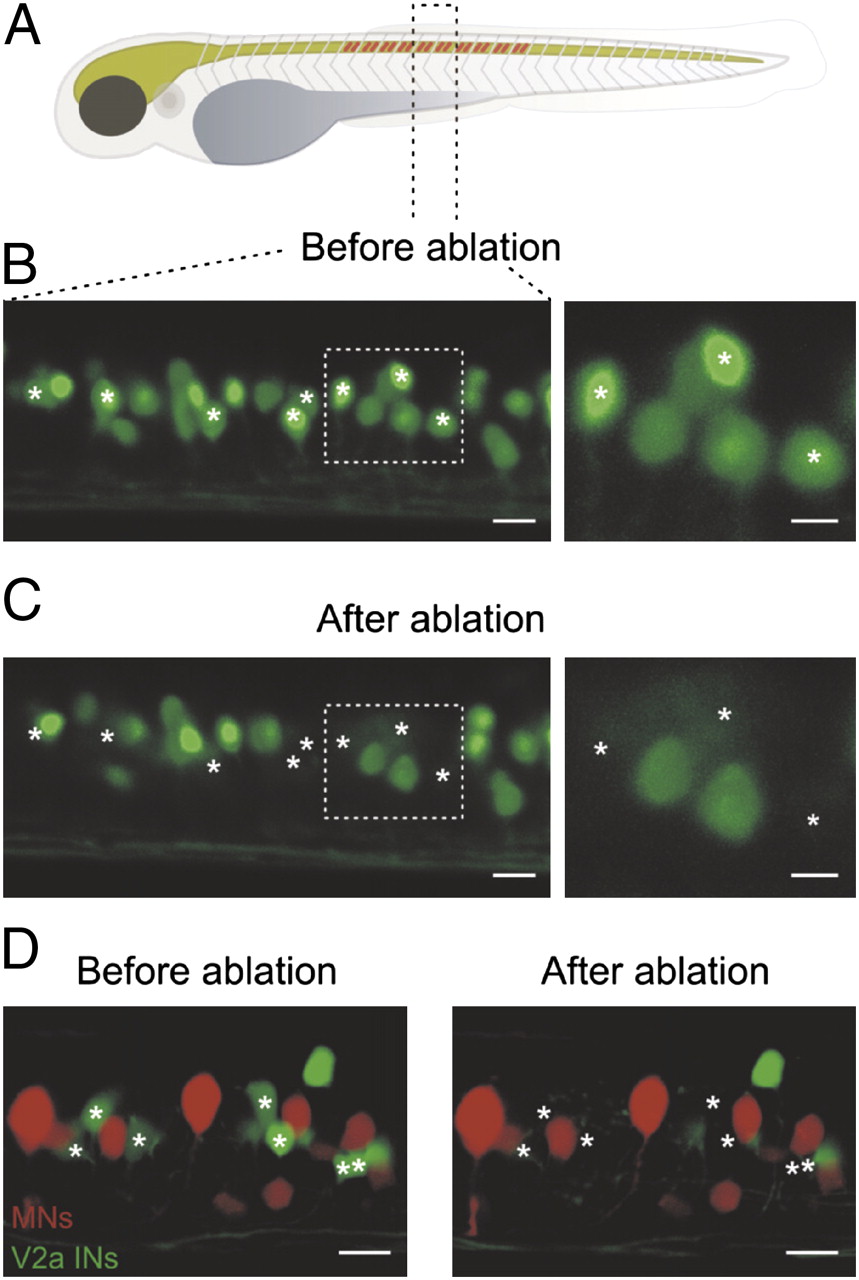
Fig. 1
Ablation of V2a interneurons in the larval zebrafish. (A) V2a interneurons were ablated over 10 segments in the midbody region of larval zebrafish. (B) Reconstruction of 1.5 segments of the spinal cord in zebrafish using confocal microscopy before the ablation of V2a interneurons. (C) The spinal cord was scanned and reconstructed after photoablation of eight V2a interneurons (asterisks). (Right) Expanded image of the area indicated by the dashed boxes. (Left scale bar, 10 μm and Right scale bar, 5 μm.) (D) Motoneurons were prelabeled with rhodamine dextran in larval zebrafish expressing GFP in V2a interneurons. Ablation of V2a interneurons did not produce any secondary damage in adjacent motoneurons. (Scale bar, 10 μm.)