
Image
|
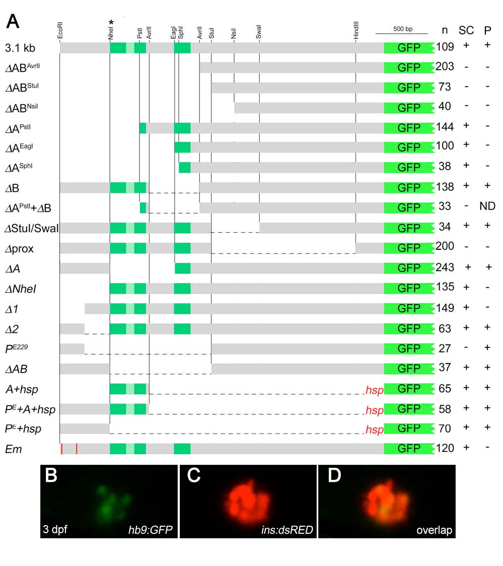
Figure Caption
Fig. S2
Characterization of 3.1 kb of upstream hb9 promoter in zebrafish
(A). Scheme of deletion constructs that were tested in transient and transgenic GFP reporter assays in 24 hpf embryos. Indicated are the number of GFP expressing embryos (n) and the presence (+) or absence (-) of GFP expression in spinal cord (SC) and pancreas (P). NheI* indicates an additional NheI site that was introduced by site-directed mutagenesis. (B-D) Confocal image projection of a Tg[ins:dsRED] embryo showing mosaic GFP expression in beta-cells following injection of hb9:GFP DNA.
Acknowledgments
This image is the copyrighted work of the attributed author or publisher, and
ZFIN has permission only to display this image to its users.
Additional permissions should be obtained from the applicable author or publisher of the image.
Reprinted from Developmental Biology, 365(1), Arkhipova, V., Wendik, B., Devos, N., Ek, O., Peers, B., and Meyer, D., Characterization and regulation of the hb9/mnx1 beta-cell progenitor specific enhancer in zebrafish, 290-302, Copyright (2012) with permission from Elsevier. Full text @ Dev. Biol.