
|
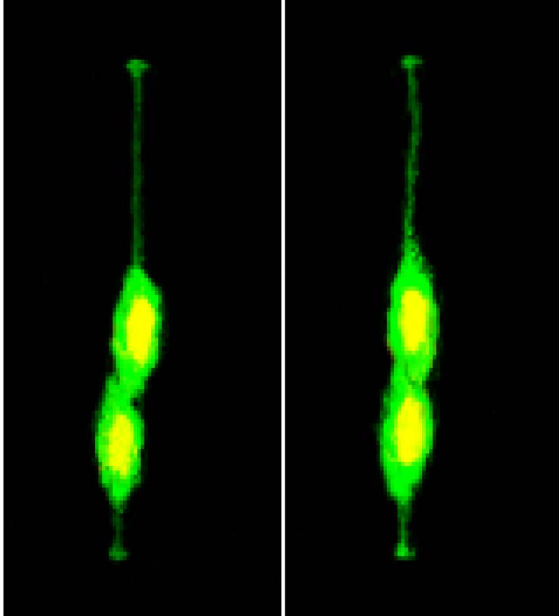
Fig. S1
Representative montage of sequential frames from in vivo time-lapse imaging of a single fluorescently-labeled mother cell in the developing zebrafish forebrain, related to Figure 1. From these images, the following can be observed: 1) 0 – 48 min, the mother cell undergoes apically directed INM to reach the apical surface. 2) 1h - 1h 24min: The mother cell divides, and its division axis is largely perpendicular to the apical basal axis. 3) 1h 36 min: The two daughter cells assume a differential positioning along the apical basal neural axis. 4) 1h 48min - 9h: The two daughter cells undergo basally directed INM while maintaining their relative apical basal positions. 5) 9h 12min – 10h 48min: The two daughter cells undergo apically directed INM while maintaining their relative apical basal positions. 6) 11h – 11h 36min: The apical daughter divides with its division axis largely perpendicular to the apical basal axis and generates two granddaughter cells that migrate basally. 7) 11h 48 min – 12h: the basal daughter divides with its division axis largely perpendicular to the apical basal axis and generates two granddaughter cells. ad: apical daughter cell which maintains a more apical position, bd: basal daughter cell which migrates to and maintains a more basal position.