
IMAGE
Fig. 2
- ID
- ZDB-IMAGE-120417-8
- Publication
- Voz et al., 2012 - Fast Homozygosity Mapping and Identification of a Zebrafish ENU-Induced Mutation by Whole-Genome Sequencing
- All Figures
- Figures for Voz et al., 2012
Image
|
Figure Caption
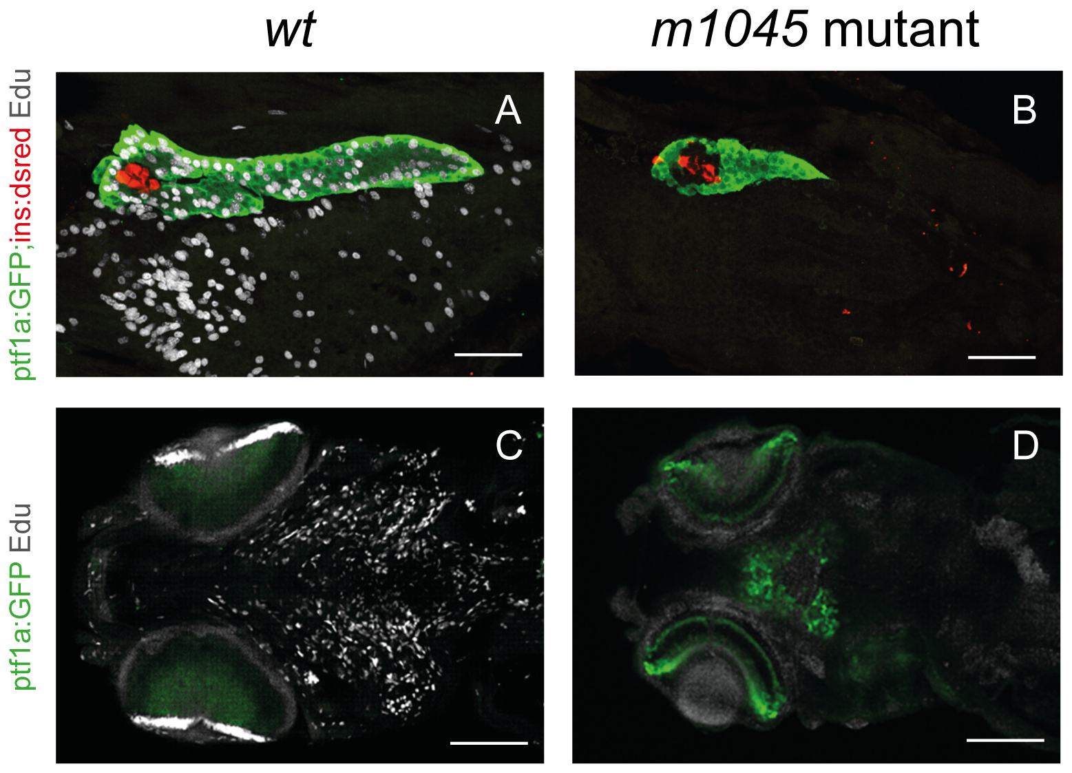
Fig. 2
The m1045 mutant displays a complete loss of cell proliferation in all tissues at 4 dpf.
1-hour Edu incorporation (95–96 hfp) of unaffected siblings (A,C) and m1045 mutant embryos (B,D) in the transgenic ptf1:GFP,ins:dsred background. Confocal projections of 4 dpf embryos of the pancreatic region (A,B) (lateral view) and of the head region (ventral view). Scale bars A,B 50 μm; C,D 100 μm. CMZ: ciliary marginal zone.
Figure Data
Acknowledgments
This image is the copyrighted work of the attributed author or publisher, and
ZFIN has permission only to display this image to its users.
Additional permissions should be obtained from the applicable author or publisher of the image.
Full text @ PLoS One