
|
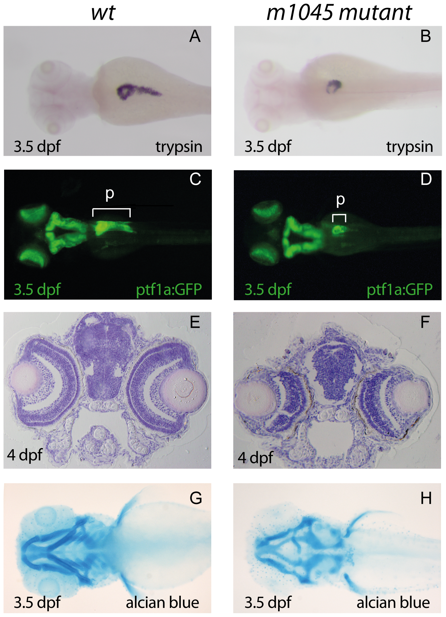
Fig. 1
The m1045 mutant exhibits hypoplasia of exocrine pancreas, eyes and branchial arches.
(A,B) : WISH using a trypsin probe of unaffected siblings (A) and m1045 mutant embryos (B) at 3.5 days post fertilization (dpf). (C–D) Dorsal view of fluorescent 3.5 dpf unaffected siblings (C) and m1045 mutants (D) in the transgenic ptf1:GFP background. (E,F) Haematoxylin/eosin staining of transverse sections of 4 dpf unaffected siblings (C) and m1045 mutants (D). Alcian blue staining of the cartilage of 3.5 dpf unaffected siblings (C) and m1045 mutants (D). A–D, G–H : views are dorsal; anterior part to the left. p: pancreas.