
|
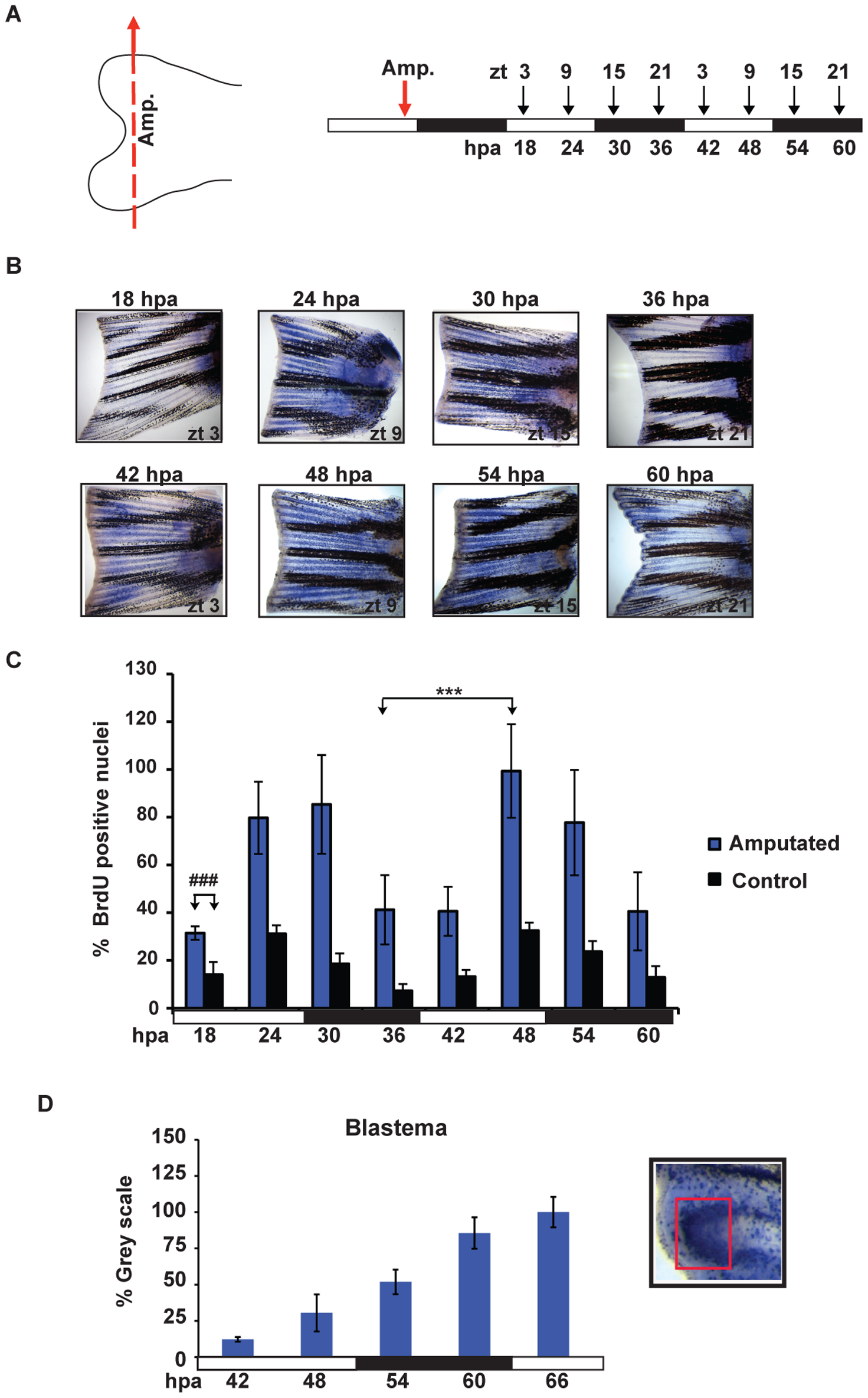
Fig. 4
Amputation differentially induces cell proliferation.
(A) Schematic representation of the experimental plan showing the amputation site (left panel), the times of sampling (ZT, right panel,,above) and the corresponding hours post amputation (hpa, right panel, below). Amputation was performed before the light-dark transition (ZT 9, red arrow) and fins were subsequently harvested every 6 hours (starting from 18 hpa). Each time point was collected following a 15 minutes incubation with BrdU. (B) Representative fins stained for BrdU incorporation at the time points indicated in panel A. (C) Quantification of the number of S-phase nuclei in amputated fins (blue bars) and non-amputated controls (black bars). On the Y-axis is plotted the % of BrdU positive nuclei with respect to the sample with the largest value (amputated, 48 hpa). The result of statistical analysis of the peak and trough values for the amputated fins is indicated by asterisks (Bonferroni′s post hoc test p<0.0001) and horizontal “brackets” above the graph. Furthermore, statistically significant differences observed at each time point between the amputated and control non-amputated fins are indicated for simplicity, by the symbol “#” and a bracket above only the first time point (18 hpa) (Bonferroni′s post hoc test p<0.0001, ###). (D) Quantification of the level of BrdU staining at the ray tips in the blastema region using Scion Image software. Inset panel (D): magnified view of the fin ray tips region stained for BrdU incorporation. A red square delimits the area that has been quantified. White and black bars below each panel indicate the light and dark periods. All the quantitative data were subjected to Cosinor analysis to test for the presence or absence of 24-h rhythmicity (see Table S1, Figure S2). Each time point represents the mean values calculated for each fin ray in a total of n = 4 to 6 fins and expressed as % of the grey scale value.