
|
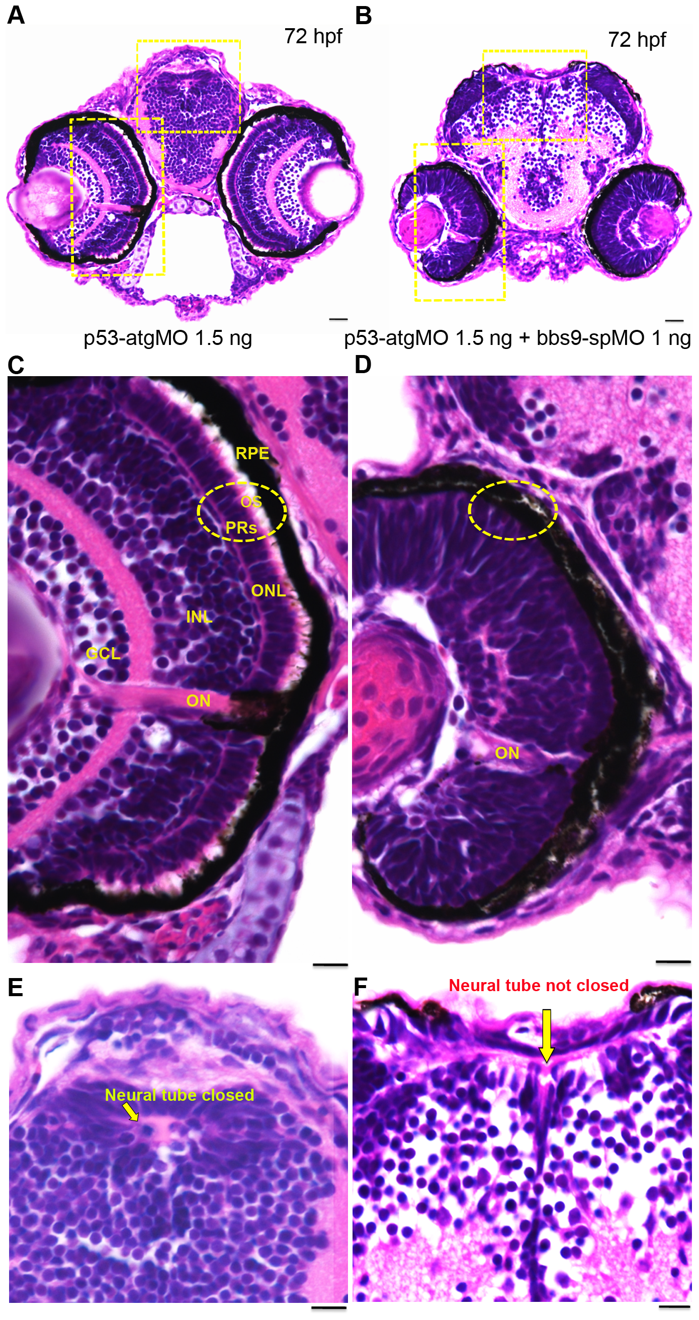
Fig. 4
bbs9-spMO morphant shows defects in retina lamination and neural tube closure.
Zebrafish head sections (72 hfp) stained with H& E. (A) The morphant injected with p53-atgMO (1.5 ng) shows normal retinal lamination and neural tube (areas highlighted with hatched yellow rectangles). (B) The morphant co-injected with bbs9-spMO (1 ng) and p53-atgMO (1.5 ng) shows lack of retinal lamination and incomplete closure of neural tube (areas highlighted with hatched yellow rectangles). (C) The morphant retina in higher magnification - area boxed in ‘A’. The retina shows all the 5 layers: Retinal pigment epithelium (RPE) abutting close to photoreceptors (PRs). The PRs (hatched circle) have visible outer segments (OS). Next to PRs are the outer nuclear layer (ONL) followed by an intact inner nuclear layer (INL) and the ganglion cell layer (GCL). The optical nerve (ON) is used as a reference point. (D) The bbs9-spMO injected morphant retina in higher magnification - area boxed in ‘B’. The retina shows no clear lamination and it lacks photoreceptor outer segments (hatched circle). (E) The morphant neural tube in higher magnification from ‘A’ shows normal closure (arrow). (F) The bbs9-spMO injected morphant neural tube in higher magnification from ‘B’ shows incomplete closure of neural tube (arrow). The scale bars indicate 100 μm.