
Fig. 9
- ID
- ZDB-IMAGE-120405-50
- Publication
- Cavaco Rodrigues et al., 2012 - Skeletal muscle regeneration in Xenopus tadpoles and zebrafish larvae
- All Figures
- Figures for Cavaco Rodrigues et al., 2012
|
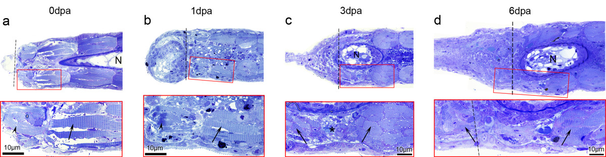
Fig. 9
Zebrafish larvae show no muscle dedifferentiation phenotype. Micrographs from semithin sections showed that larval tail myofibres have a well organized sarcomeric structure that do not show signs of dedifferentiation during regeneration (arrows). (a, b) At 0 dpa and 1 dpa, distal most fibres show altered sarcomeric striations, characteristic of damaged or dying myofibre (arrowheads). (c) At 3 dpa this phenotype was not observed (neither at 2 dpa, not shown), instead, less muscle is observed close to the amputation plane (asterisk). (d) At 6 dpa, new myofibres are visible in the regenerate (left arrow). Notochord (N) is visible as an oval at 3 and 6 dpa because of the slight dorsal curvature that regenerating tails acquire (see Figure 8h). Dashed lines: amputation planes. n e four sections per animal, three animals per time point.