
|
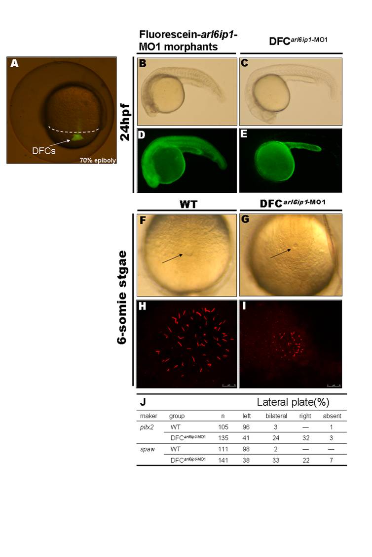
Fig. S7 Specific knockdown of Arl6ip1 in DFCs alters LR development without other effects on embryogenesis. (A) As observed under fluorescent microscopy, injection of fluorescein-tagged arl6ip1-MO1 into mid-blastula stage embryos was observed in dorsal forerunner cells (DFCs) 3–4 hr post-injection at 70% epiboly stage. The dorsal margin was indicated by dashed line. (B–E) Lateral views of 24hpf embryos. Embryos were observed under transmitted light microscopy (B, C) or fluorescent microscopy (D, E). MOs were present in all cells of embryos injected at the one-cell stage (D), and severe defects induced by fluorescein-tagged arl6ip1-MO1 embryos were shown (B). Fluorescein-tagged arl6ip1-MO1 injected into embryos at mid-blastula stage was primarily found in the yolk cell and yolk tube (E), indicating that cells, other than DFC and yolk, did not incorporate MO. These DFC of arl6ip1-MO1-injected embryos (DFCarl6ip1-MO1) developed a normal morphology (C) similar to WT embryos. (F, G) Tail views of 6-somite stage embryos. KV was indicated by arrows. Compared to WT embryos (F), DFCarl6ip1-MO1 embryos showed reduced KV at 6-somite stage (G). (H, I) Confocal microscopy images of anti-acetylated tubulin staining of KV cilia (red fluorescent) at 6-somite stage. Cilia distributed over a spherical pattern in the region of KV (H). However, cilia were disorganized in reduced KV of DFCarl6ip1-MO1 embryos (I). (K) Summary of pitx2 and spaw asymmetrical gene expression patterns.