
|
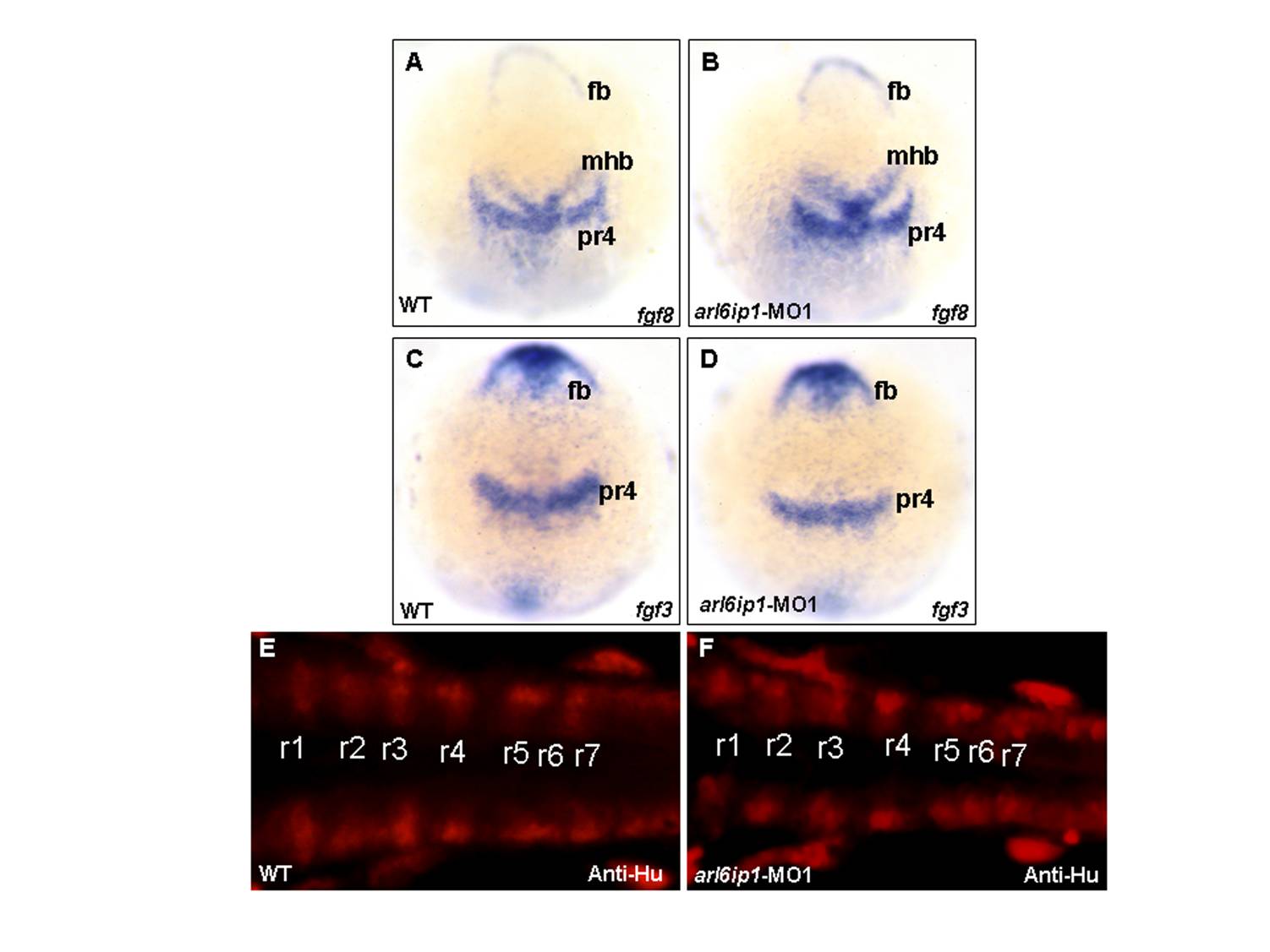
Fig. S2 The arl6ip1-MO1-injected embryos do not appear to have defective patterning in hindbrain. Wild-type (WT; A, C, E) and arl6ip1-knockdown (MO; B, D, F) embryos, either at 3-somite stage (3ss) (A-D) or at 24 hpf (E, F), were observed at dorsal view. (A-D) Neither fgf3 expression nor fgf8 expression in the arl6ip1 morphants was distinguishable from that of wild-type embryos (A vs. B; C vs. D). (E, F) Dorsal views of 24 hpf embryos processed for anti-Hu immunofluorescence staining (IFA) to reveal hindbrain segmentation. Similar to WT embryos, arl6ip1-knockdown embryos showed normal r1-r7 segmentation. fb, forebrain; mhb, midbrain-hindbrain border; pr4, premature rhombomere 4; r1–r7, rhombomere 1–7.