
|
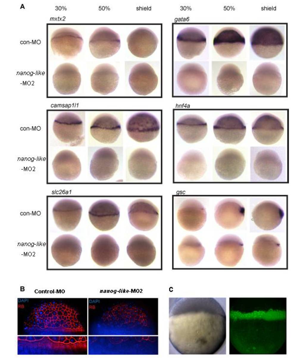
Fig. S4 Lack of YSL transcription in nanog-like morphants, related to Figure 3
(A) The expression of gata6, mxtx2, camsap1l1, hnf4a, and slc26a1 is absent in the YSL of nanog-like morphants. The shield structure is formed in nanog-like morphants, as indicated by the expression of gsc. All views are lateral with the presumptive dorsal side to the right.
(B) The F-actin ring is absent in nanog-like morphants. F-factin was stained with rhodamine phalloidin (RP). Nuclei were stained with DAPI. White brackets indicate the F-actin ring in the YSL.
(C) Injection of mRNA into the yolk of 2.5 hpf embryos enables YSL-specific expression. GFP mRNA was injected into the yolk of a 2.5 hpf embryo. The embryo was imaged at the dome stage. Left: white field; right: GFP channel.
Reprinted from Developmental Cell, 22(3), Xu, C., Fan, Z.P., Müller, P., Fogley, R., Dibiase, A., Trompouki, E., Unternaehrer, J., Xiong, F., Torregroza, I., Evans, T., Megason, S.G., Daley, G.Q., Schier, A.F., Young, R.A., and Zon, L.I., Nanog-like Regulates Endoderm Formation through the Mxtx2-Nodal Pathway, 625-238, Copyright (2012) with permission from Elsevier. Full text @ Dev. Cell