
IMAGE
Fig. S4
- ID
- ZDB-IMAGE-120329-16
- Publication
- Dempsey et al., 2012 - PhOTO Zebrafish: A Transgenic Resource for In Vivo Lineage Tracing during Development and Regeneration
- All Figures
- Figures for Dempsey et al., 2012
Image
|
Figure Caption
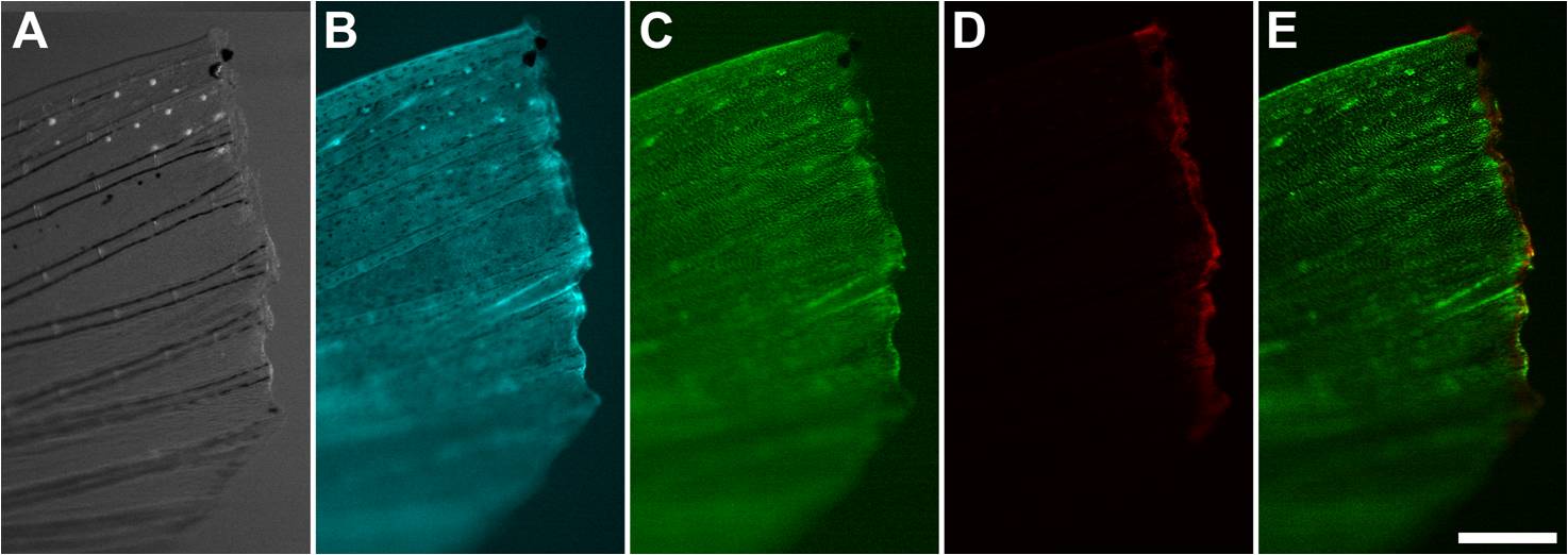
Fig. S4 Amputated Adult PhOTO-N Zebrafish Tail Fin After Photoconversion. A fluorescent stereomicroscope image of the photoconverted amputated tail fin (anterior left, ventral down) showing (A) bright field, (B) memb-Cerulean (blue), (C) unconverted H2B-Dendra2 (green), (D) photoconverted H2B-Dendra2 (red), and (E) a merged image of (C) and (D). Note that almost all of the H2B-Dendra2 fluorescence has been photoconverted in the ~50–100μm region along the amputation plane, as is indicated by the lack of green signal in the photoconverted stripe in (C). Scale bar is ~300μm.
Acknowledgments
This image is the copyrighted work of the attributed author or publisher, and
ZFIN has permission only to display this image to its users.
Additional permissions should be obtained from the applicable author or publisher of the image.
Full text @ PLoS One