
Fig. 4
- ID
- ZDB-IMAGE-120329-12
- Publication
- Dempsey et al., 2012 - PhOTO Zebrafish: A Transgenic Resource for In Vivo Lineage Tracing during Development and Regeneration
- All Figures
- Figures for Dempsey et al., 2012
|
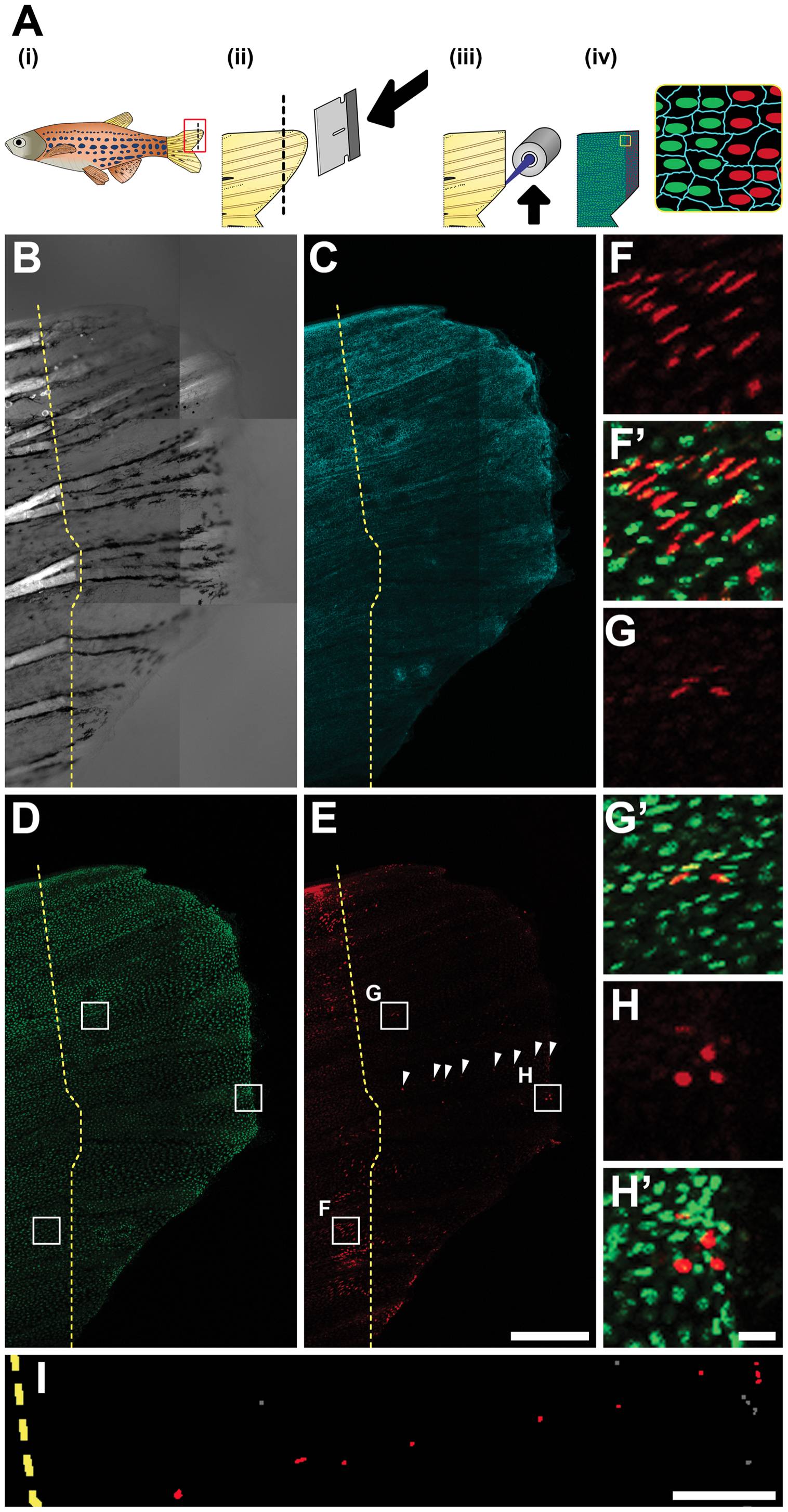
Fig. 4 Photoconverted Cell Organization After Epimorphic Tail Fin Regeneration in a Living Adult PhOTO-N Zebrafish.
(A) Overview of fin regeneration protocol. (i) An adult zebrafish is anesthetized and (ii) the tail is amputated using a razor blade (dotted line refers to amputation plane). (iii) Photoconversion is achieved by fluorescent illumination along the amputation plane. (iv) In addition to the H2B-Dendra2 (green) and memb-Cerulean (blue) signal, there is now a line of photoconverted H2B-Dendra2 nuclei (red). (B-I) Lateral view (anterior left, ventral down) MIP images of the regenerating tail fin of a live, anesthetized adult zebrafish 7 days post amputation. The dotted yellow line indicates the approximate amputation plane. (B) Bright field channel. Bright signal to the left of the dotted line arises from tissue birefringence. The terminal end of this signal indicates the amputation plane. (C) Memb-Cerulean (blue) and (D) unconverted H2B-Dendra2 (green) channels indicate that expression is still consistent as the tail fin regenerates. (E) Many of cells with fluorescence above background appear both before and after the plane of amputation in the photoconverted H2B-Dendra2 (red) channel. Cells in the central region of the tail show the least expression, suggesting extensive cell division contributing to regeneration in this area. However, certain cells to the right of the amputation plane are bright, aligned, and evenly spaced (arrowheads). (F-H) Zoomed boxed regions from (D) and (E) of photoconverted H2B-Dendra2 (red) alone. (F′-H′) Merged unconverted H2B-Dendra2 (green) and photoconverted H2B-Dendra2 (red) within the zoomed boxed regions from (D), (E). (F, F′) Many photoconverted cells in the ventral and dorsal portions of the amputated fin stayed behind the amputation plane during the regeneration process. (G, G′) Surprisingly, a subset of brightly photoconverted cells were found in the central portion in addition to (H, H′) other bright cells at the distal edge of the regenerate. (I) Zoomed binary image of a subset of the segmented nuclei from the photoconverted H2B-Dendra2 channel in panel (E). Among the scattered cells with photoconverted signal over background (gray), certain cells (red) seem aligned and evenly spaced along the anterior-posterior axis of the tail fin within the regenerating region. Scale bar for (B-E) is 300μm. Scale bar for (F-H) is ~10μm. Scale bar for (I) is ~100μm.