
|
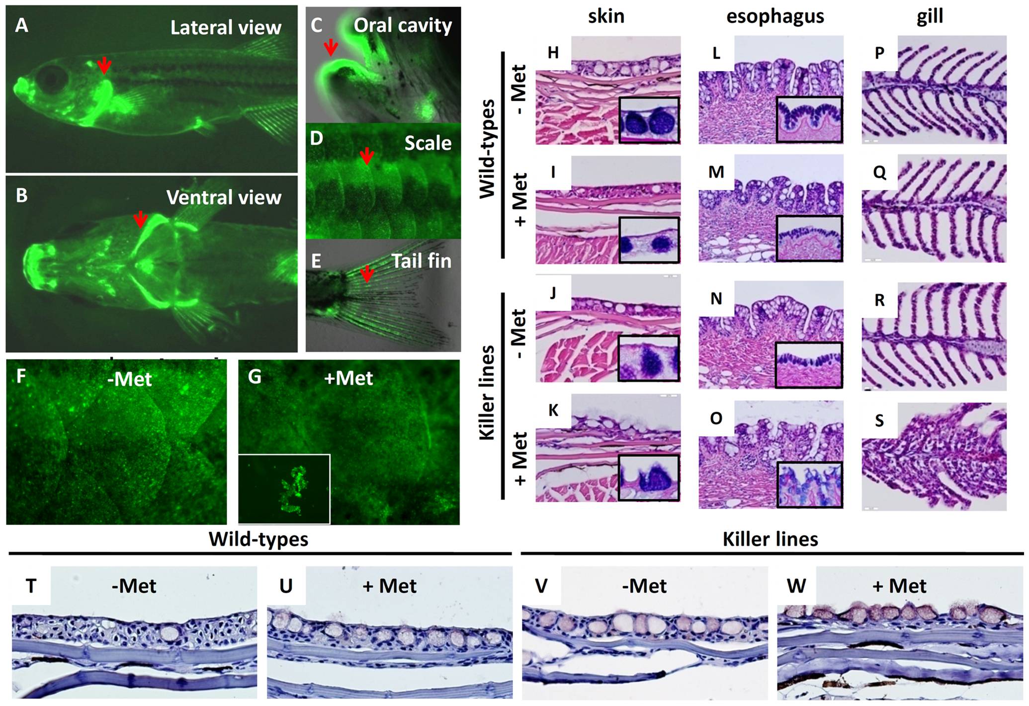
Fig. 5 Evaluation of skin ablation by NTR/Met ablation system in adult killer line.
The lateral (A) and ventral (B) views of fluorescent appearance of NTR-hKikGR fusion protein in the killer line at adult stage. Some regions like gill operculum (A, B), oral cavity (C), scale (D) and tail fin (E), which are rich in epithelial structures, showed robust fluorescent signals (heighted by arrows). (F–G) Test of the possibility of performing skin ablation in the adult killer line. Treatment of the killer line with 2.5 mM Met for three consecutive days resulted in greatly compromised skin integrity and some detached skin debris (NTR-hKikGR+) in the fish tank. (H–S) Histological assessment of skin integrity in wild-types or killer lines treated with or without 2.5 mM Met. Paraffin sections stained with hematoxylin and eosin showing the serial morphological changes in the regions of superficial skin (H–K), esophagus (L–O) and gill epithelium (P–S). Included for comparison, the normal epidermal histology in superficial skin (H), esophagus (L) and gill (P) before performing skin ablation. To highlight the position of mucous cells, PAS staining (blue color) in paraffin sections derived from skin (H–K) and esophagus (L–O) shown in lower right corners. (T–W) Detection of cell apoptosis in the damaged skin by activated caspase 3 antibody staining on paraffin sections (brown signals). The killer line adults were first incubated with 2.5 mM Met solution for three consecutive days to execute cell ablation and then paraffin sections, at 5 μm intervals, were cut for histological assay or immunohistochemistry. Met, metrodinazole; PAS, Periodic Acid Schiff.