
|
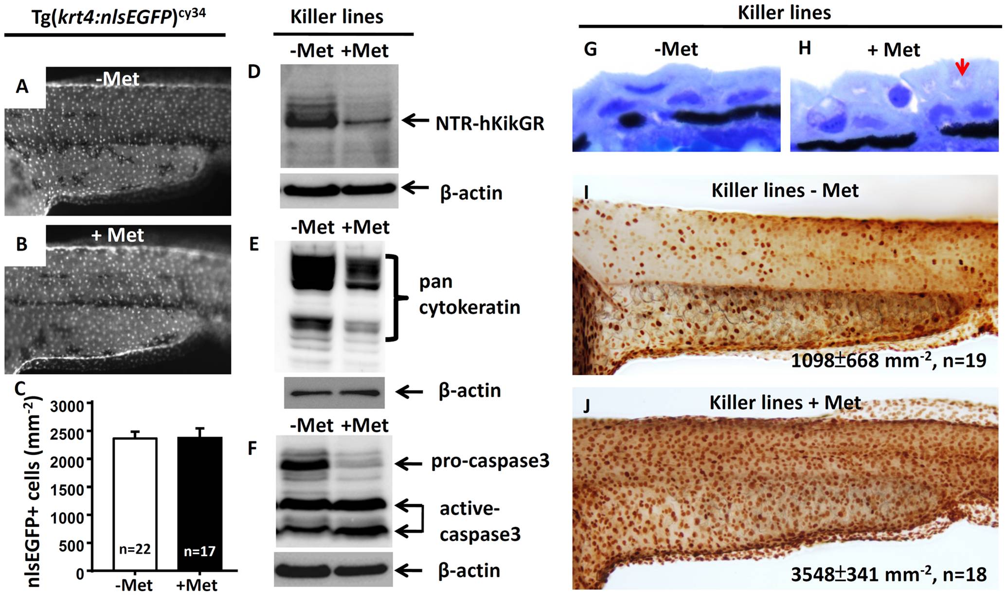
Fig. 4 Apoptotic cell death mediates the loss of NTR-hKikGR+ fluorescent signals in Met-treated killer line.
krt4 promoter suppression does not mediate loss of NTR-hKikGR+ fluorescence in Met-treated killer line because downregulation of nlsEGFP+ fluorescence signal intensity (A and B) or nlsEGFP+ cells (C) in Tg(krt4:nlsEGFP)cy34 did not occur with or without Met treatment. (D–F) Western blot analysis showed that apoptotic cell death mediates the loss of NTR-hKikGR+ fluorescent signals in Met-treated killer line embryos because the relative expression levels of NTR-hKikGR fusion protein (D), pan-cytokeratin (E) and pro-caspase 3 (F) greatly reduced in Met-treated embryos. Note that the relative intensities of the cleavaged caspase 3-immunoreactive signals greatly increased in the Met-treated killer line (F). (G–H) Plastic sections at 2 µm thickness show the greatly compromised superficial skin integrity in Met-treated killer line embryos (arrow). Whole-mount TUNEL assay demonstrated significantly enhanced cell death signals in Met treated killer line embryos (J) compared to the untreated group (I). The cell number is presented as the mean±S.D. Student′s t-test was used to make statistical comparisons between untreated (-Met) and Met-treated (+Met) killer lines. Met, metrodinazole.