
|
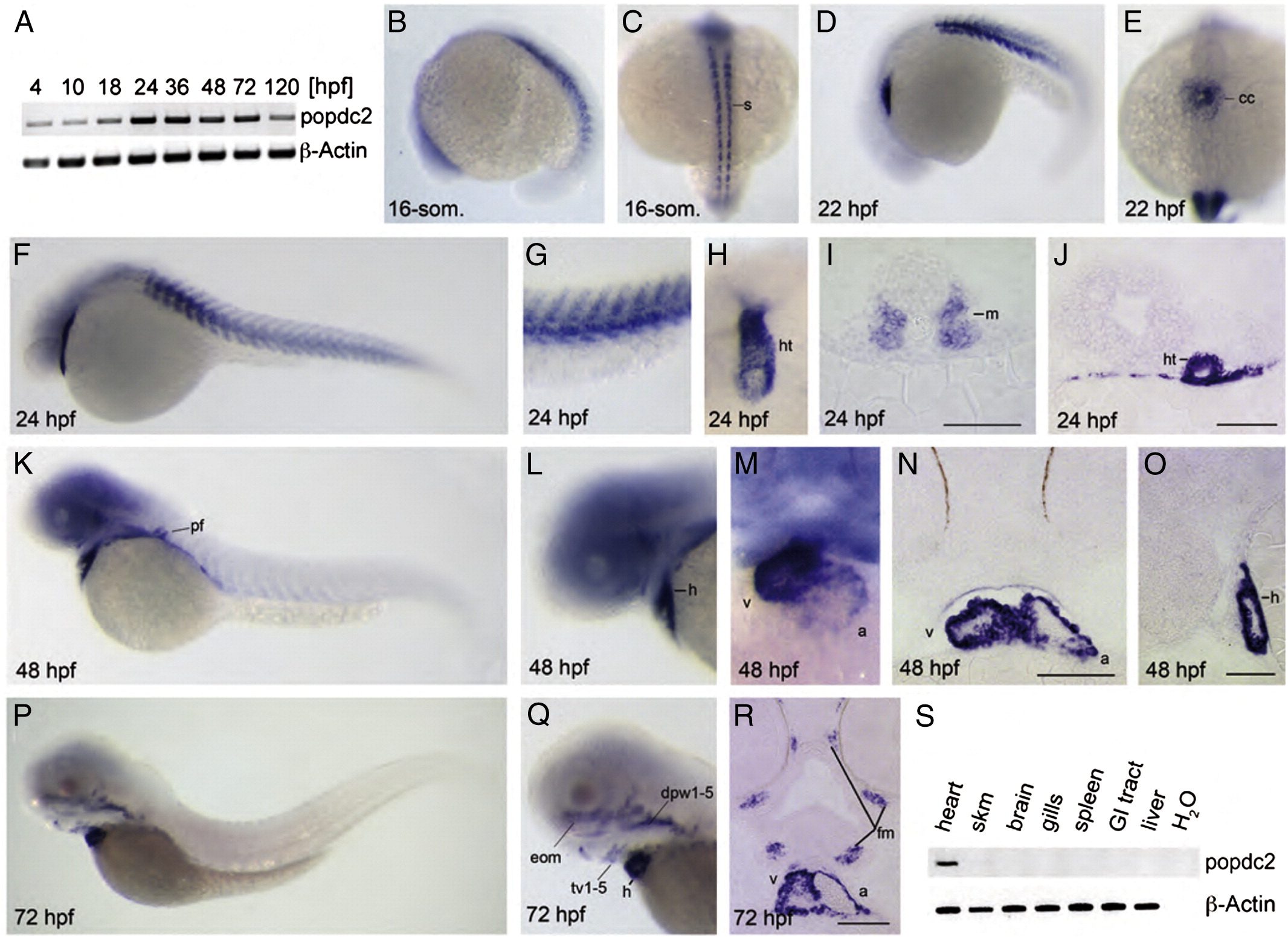
Fig. 1 Expression of popdc2 during zebrafish development. (A) RT-PCR analysis of popdc2 expression during development. (B–R) Whole mount in situ hybridization analysis using a full-length popdc2 probe of zebrafish embryos at (B,C) 16-somites stage, (D,E) 22 hpf, (F–H) 24 hpf, (K–M) 48 hpf, and (P–Q) 72 hpf. Stained embryos are viewed from (B,D,F,G,K,L,P,Q) left lateral, (C,E) dorsal, and (H,M) ventrofrontal. Panel (I,J,N,R) depicts transversal and (O) sagittal sections through stained embryos. (S) RT-PCR analysis of popdc2 expression in adult tissues. Abbreviations: a—atrium; cc—cardiac conus; dpw1–5—dorsal pharyngeal wall muscles 1–5; eom—extraocular muscles; fm—facial muscles; GI tract—gastro-intestinal tract; h—heart; ht—tubular heart; m—myotome; pf—pectoral fin bud; s—somites; skm—skeletal muscle; tv1–5—transversus ventralis muscles; v—ventricle. Scale bar = 100 μm.
Reprinted from Developmental Biology, 363(2), Kirchmaier, B.C., Poon, K.L., Schwerte, T., Huisken, J., Winkler, C., Jungblut, B., Stainier, D.Y., and Brand, T., The Popeye domain containing 2 (popdc2) gene in zebrafish is required for heart and skeletal muscle development, 438-450, Copyright (2012) with permission from Elsevier. Full text @ Dev. Biol.