
Image
|
Figure Caption
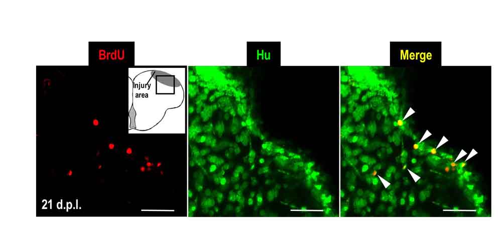
Fig. S3 Newborn cells differentiate into Hu-positive neurons in the injury site. Immunodetection of BrdU (red) and Hu protein (green) in coronal brain sections (dorsal up) of injured adult wild-type zebrafish at 21 d.p.l.. White arrowheads indicate BrdU and Hu double-positive cells. BrdU-labeling protocol: see Fig. 4A. Scale bars: 50 μm.
Acknowledgments
This image is the copyrighted work of the attributed author or publisher, and
ZFIN has permission only to display this image to its users.
Additional permissions should be obtained from the applicable author or publisher of the image.
Full text @ Dis. Model. Mech.