
|
Fig. S1
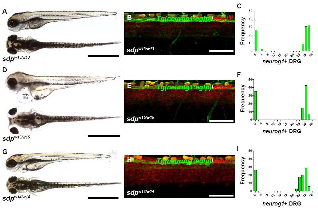
All alleles of sdp exhibit complete DRG loss. (A) sdpw13/w13 exhibits normal morphology and pigmentation. Scale bar: 500 μm. (B) A high-magnification image of 3 dpf Tg(neurog1:egfp)/sdpw13/w13 zebrafish immunostained for Elavl1 reveals no DRG neurons. Scale bar: 50 μm. (C) Approximately 25% of 3 dpf embryos from a cross between sdpw13/+ parents fail to form neurog1+ DRG, indicating that sdpw13 is a fully penetrant recessive mutation. (D) sdpw15/w15 exhibits normal pigmentation but acquires a progressively worsening edema around the eyes and heart beginning at 3 dpf. Scale bar: 500 μm. (E) A high-magnification image of 3 dpf Tg(neurog1:egfp)/sdpw15/w15 zebrafish immunostained for Elavl1 reveals no DRG neurons. Scale bar: 50 μm. (F) Approximately 25% of 3 dpf embryos from a cross between sdpw15/+ parents fail to form neurog1+ DRG, indicating that sdpw15 is a fully penetrant recessive mutation. (G) sdpw14/w14 exhibits normal morphology and pigmentation. Scale bar: 500 μm. (H) A high-magnification image of 3 dpf Tg(neurog1:egfp)/sdpw14/w14 zebrafish immunostained for Elavl1 reveals no DRG neurons. Scale bar: 50 μm. (I) Approximately 25% of 3 dpf embryos from a cross between sdpw14/+ parents fail to form neurog1+ DRG, indicating that sdpw14 is a fully penetrant recessive mutation.