
Image
|
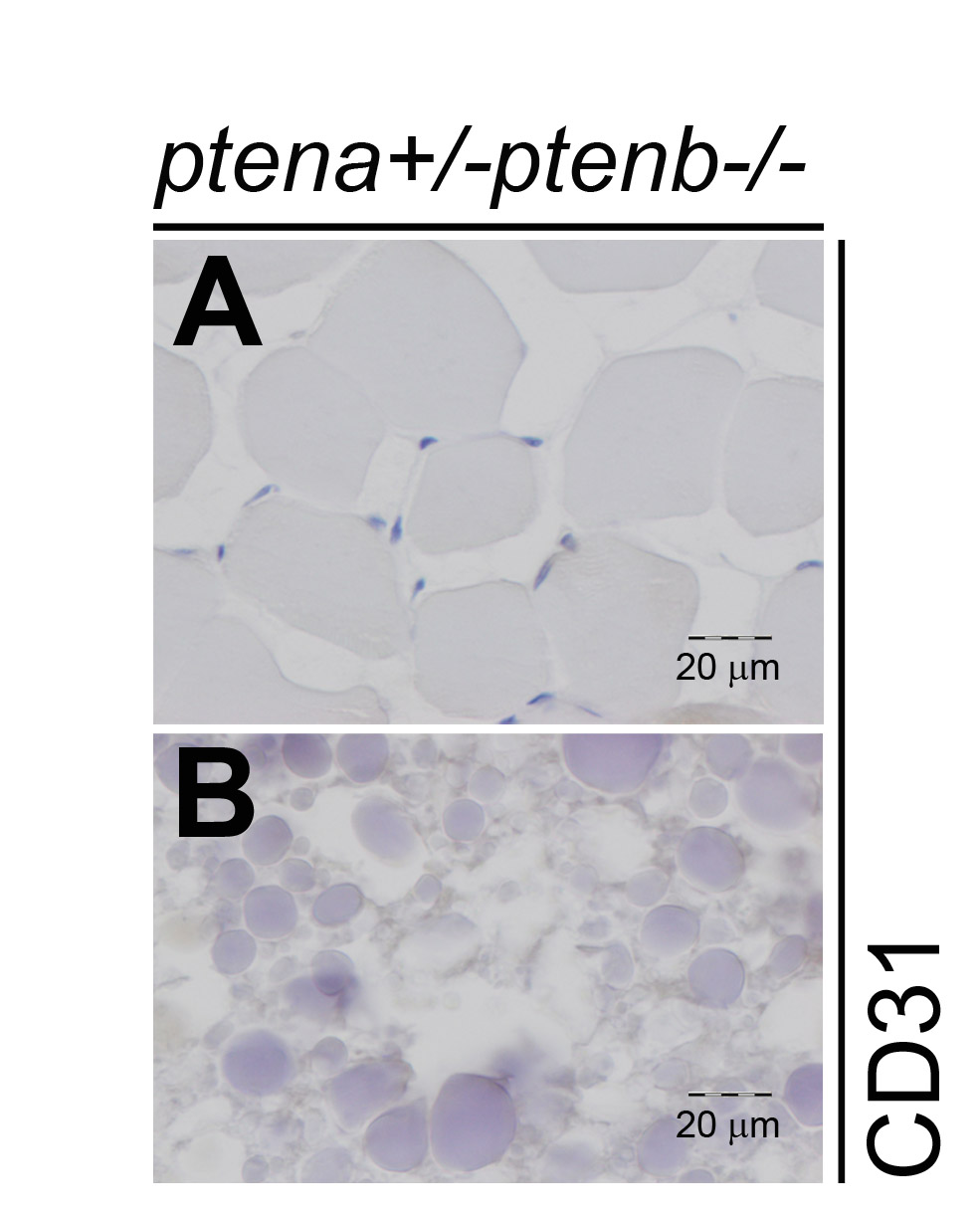
Figure Caption
Fig. S1
Muscle and ovary tissue do not exhibit CD31 staining. Immunohistochemistry on transversal sections from a ptena+/-ptenb-/- mutant fish with a diagnosed hemangiosarcoma, using CD31 specific antibody. Different areas from the CD31-stained sections in Fig. 4 are depicted here, which contain (a) muscle and (b) ovary. Both do not show CD31 staining, as expected for these non-endothelial tissues.. Sections of representative tissues are depicted. Scale bar is 20 μm, as indicated.
Acknowledgments
This image is the copyrighted work of the attributed author or publisher, and
ZFIN has permission only to display this image to its users.
Additional permissions should be obtained from the applicable author or publisher of the image.
Full text @ Dis. Model. Mech.