
|
Fig. 10
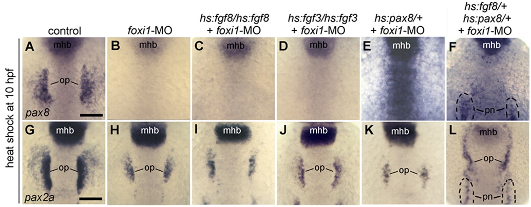
Misexpression of Fgf or Pax8 cannot expand otic development without Foxi1. (A–F) Expression of pax8 at 13 hpf. (G–L) Expression of pax2a at 13 hpf. Embryos were heat shocked at 10 hpf, and most embryos were injected at the one-cell stage with foxi1-MO (B–F, H–L) as indicated across the top of the figure. Genotypes of transgenic embryos, including hs:fgf8/hs:fgf8 (C, I), hs:fgf3/hs:fgf3 (D, J), hs:pax8/+ (E, K), and hs:fgf8/+; hs:pax8/+ (F, L) are indicated across the top of the figure. Positions of midbrain–hindbrain boundary (mhb), pronerphros (pn) and otic placode (op) are indicated. Note that the pronerphric domain of pax2a is also expanded anteriorly in double transgenic embryos (L). All images show dorsal views with anterior to the top. Scale bar, 150 μm.
Reprinted from Developmental Biology, 364(1), Padanad, M.S., Bhat, N., Guo, B., and Riley, B.B., Conditions that influence the response to Fgf during otic placode induction, 1-10, Copyright (2012) with permission from Elsevier. Full text @ Dev. Biol.