
|
Fig. 4
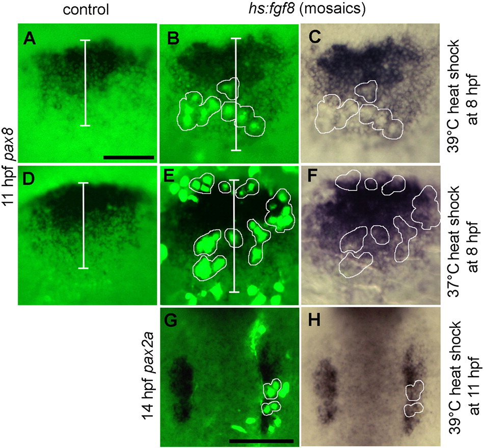
Effects of mosaic misexpression of Fgf8. (A–F) Lateral views (anterior to the left) showing expression of pax8 at 11 hpf in the otic domain in control embryos (A, D) or hs:fgf8/+ mosaic embryos (B, C, E, F) heat shocked at 39 °C (A–C) or 37 °C (D–F) at 8 hpf. Clusters of transgenic cells (green) are encircled with white borders to facilitate comparison of fluorescent images (B, E) with bright field images of the same specimens (C, F). White bars (A, B, D and E) mark the ML width of the otic domain. Note that transgenic cells express pax8 following heat shock at 37 °C but not at 39 °C, yet the otic domain is laterally expanded at both temperatures. (G, H) Dorsal views (anterior to the top) showing expression of pax2a at 14 hpf in a mosaic embryo heat shocked at 39 °C at 11 hpf. Transgenic cells (green, with white borders) express pax2a and the otic domain is lengthened along the AP axis. Scale bar, 150 μm.
Reprinted from Developmental Biology, 364(1), Padanad, M.S., Bhat, N., Guo, B., and Riley, B.B., Conditions that influence the response to Fgf during otic placode induction, 1-10, Copyright (2012) with permission from Elsevier. Full text @ Dev. Biol.