
|
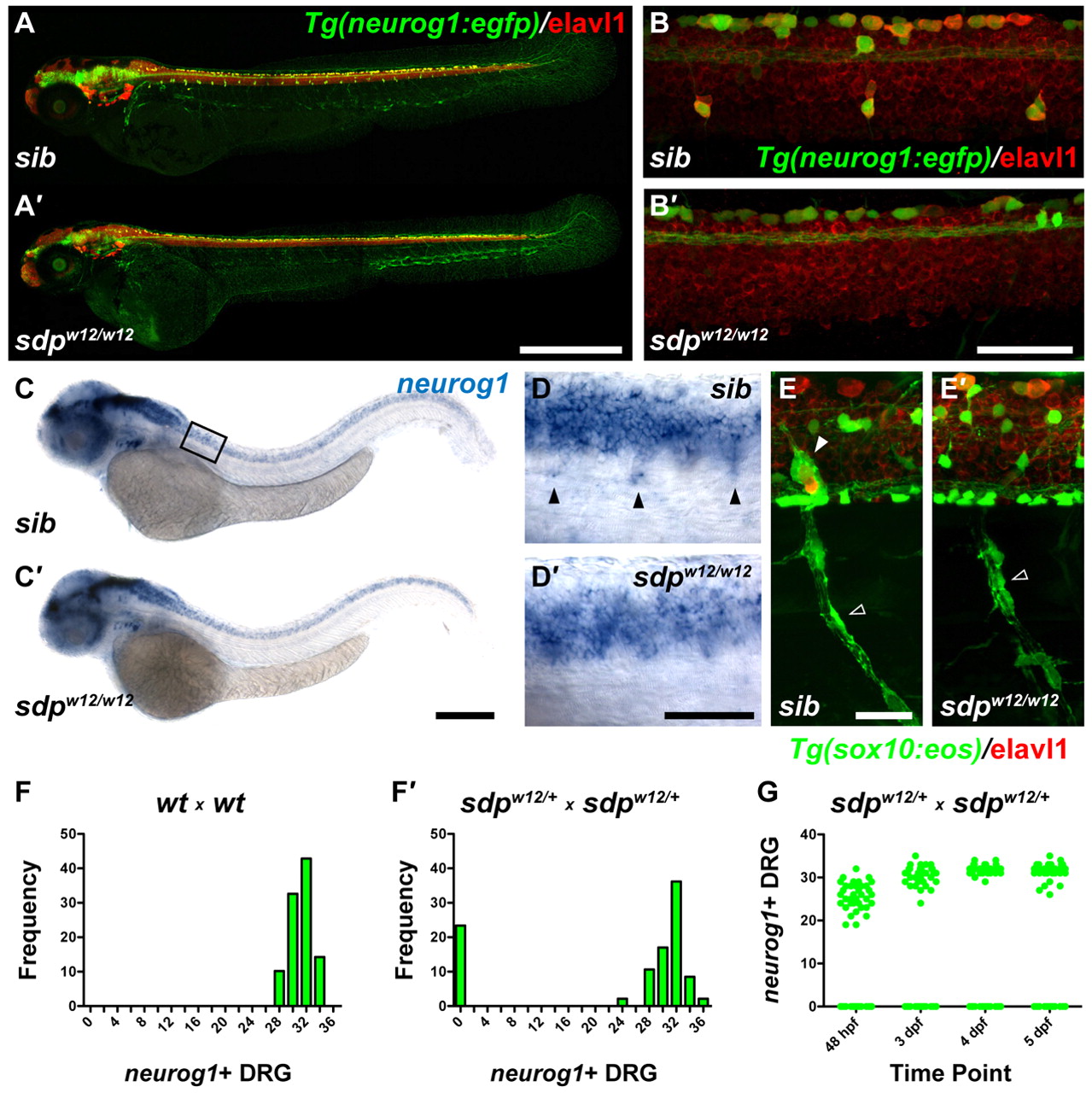
Fig. 1
sensory-deprived (sdp) mutants exhibit a complete loss of DRG sensory neurons. (A) Tg(neurog1:egfp) zebrafish embryos (3 dpf) immunostained for Elavl1. (A2) DRG are absent in an sdpw12/w12 mutant embryo. Scale bar: 500 μm. (B,B2) High magnification images of A and A2. Scale bar: 50 μm. (C,C2) neurog1 expression at 48 hpf. Box indicates position of high magnification images in D,D2. Scale bar: 250 μm. (D,D2) High magnification image of C and C2. Scale bar: 50 μm. Arrowheads indicate neurog+ DRG sensory neuron precursors. (E,E2) Tg(sox10:eos) embryo (3 dpf) immunostained for Elavl1. Although Schwann glia (empty arrowhead) are retained, satellite glia (filled arrowhead) are absent in sdpw12/w12. Scale bar: 25 μm. (F,F2) Counts of neurog1+ DRG at 3 dpf. Approximately 25% of embryos fail to form DRG, suggesting that sdpw12 is a fully penetrant recessive mutation. (G) Counts of neurog1+ DRG followed over four days. DRG never appear in <25% of the population, indicating that sdp does not delay in DRG development.