
|
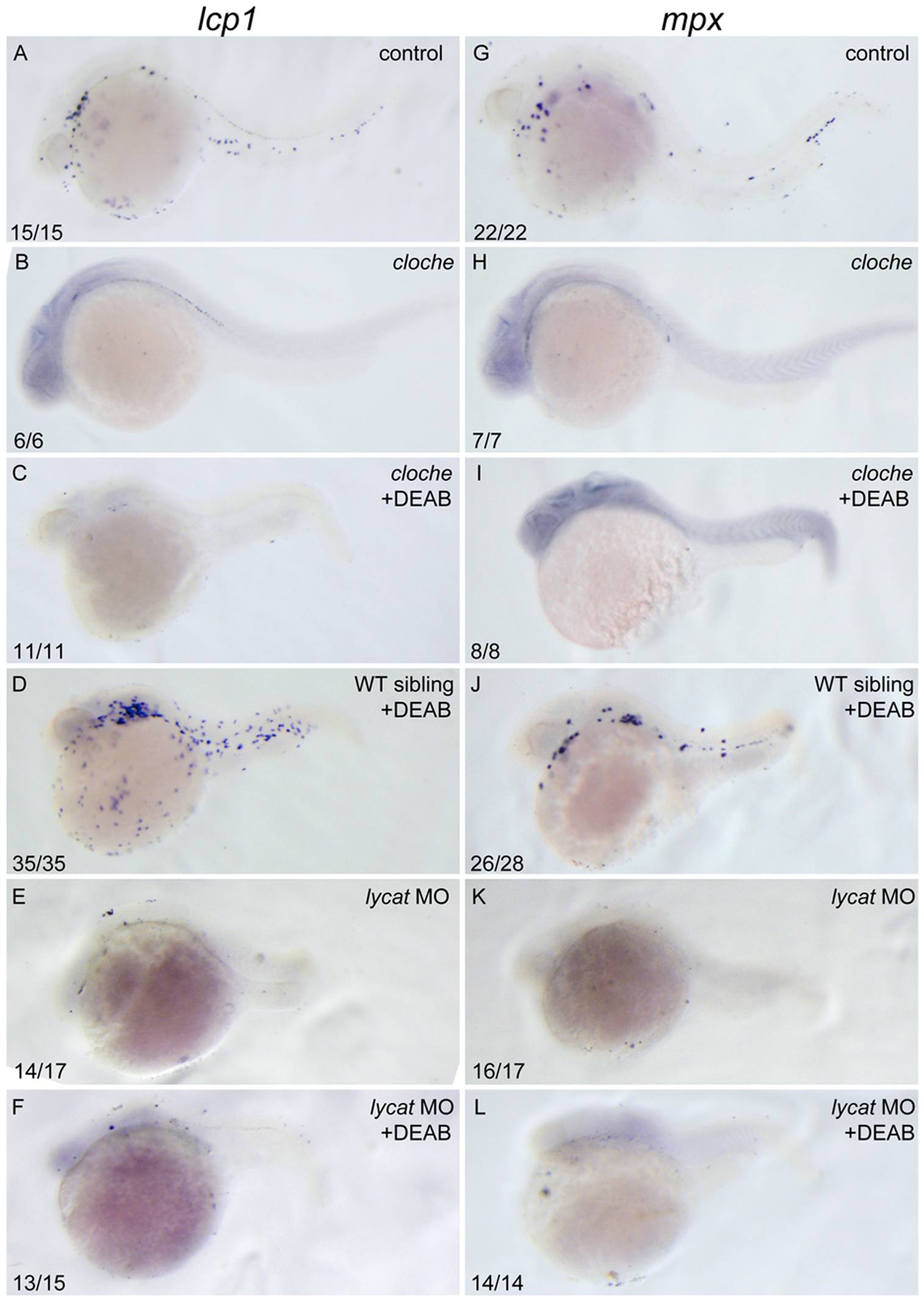
Fig. 6 DEAB cannot rescue the defective primitive myelopoiesis in cloche or lycat knockdown embryos.
All embryos are positioned anterior left and lateral front. Wild type siblings (A, G), cloche (B, H) and the embryos microinjected with lycat-MO at 1–2-cell stage (E, K) were treated with vehicle DMSO whereas cloche (C, I), cloche siblings (D, J) and lycat-MO knockdown (F, L) embryos were treated with 10 μM DEAB from 1–2-cell stage until 26 hpf. They were then examined for expressions of myeloid markers lcp1 (A–F) and mpx (G–L) at 26 hpf by whole mount in situ hybridization. The number shown in the lower left-hand corner of each panel is the number of embryos exhibiting the typical phenotype shown in the panel to the number of embryos totally observed.