
|
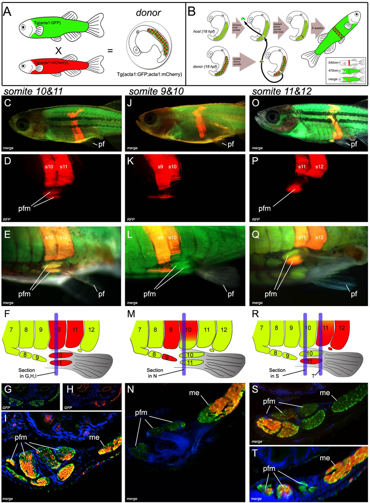
Fig. 5 Transgenic somite transplantation in D. rerio reveals pelvic fin muscles derive from myotomal extension.
(A) Donor embryos are generated by crossing Tg(acta1:mCherry)pc4 with Tg(acta1:GFP)zf13 [33]. (B) Donor somites are surgically removed and transplanted into the host. (C–I) Myotomal extensions derived from somites 10 and 11 generate the pelvic fin muscles (Section through s10 (blue line in F) of host (5 wk post-operation) (G, H, and I)). (Comprehensive methods are provided in supplementary data, however surgery involving transplantation of two consecutive somites lead to a greater probability of transplanting the entire somite, including the ventral aspect required for pelvic fin muscle formation). (J–L) Contribution to the pelvic fin muscle requires the most ventral tip of the somite to be transplanted. Full transplantation of somite 9 contributes to ventral muscle anterior to pelvic fin. but a partial transplant of somite 10 does not result in contribution to the pelvic fin muscle. (M, N) Section through s10 (blue line in M) of the transplanted host (5 wk post-operation). (N) If the donor somite is not included in the most ventral tip of the extension, contribution to the fin does not occur . (O–T) The most ventral group of pelvic fin muscles are derived from somite 11 (s11+s12) section through s10 (S) (blue line in R) and s11 (T) (blue line in R) of host (5 wk post-operation). Pf, pelvic fin; rfp, red fluorescent protein; pfm, pelvic fin muscle; me, myotomal extension; s9,s10,s11,s12, somite numbered from anterior to posterior.