
|
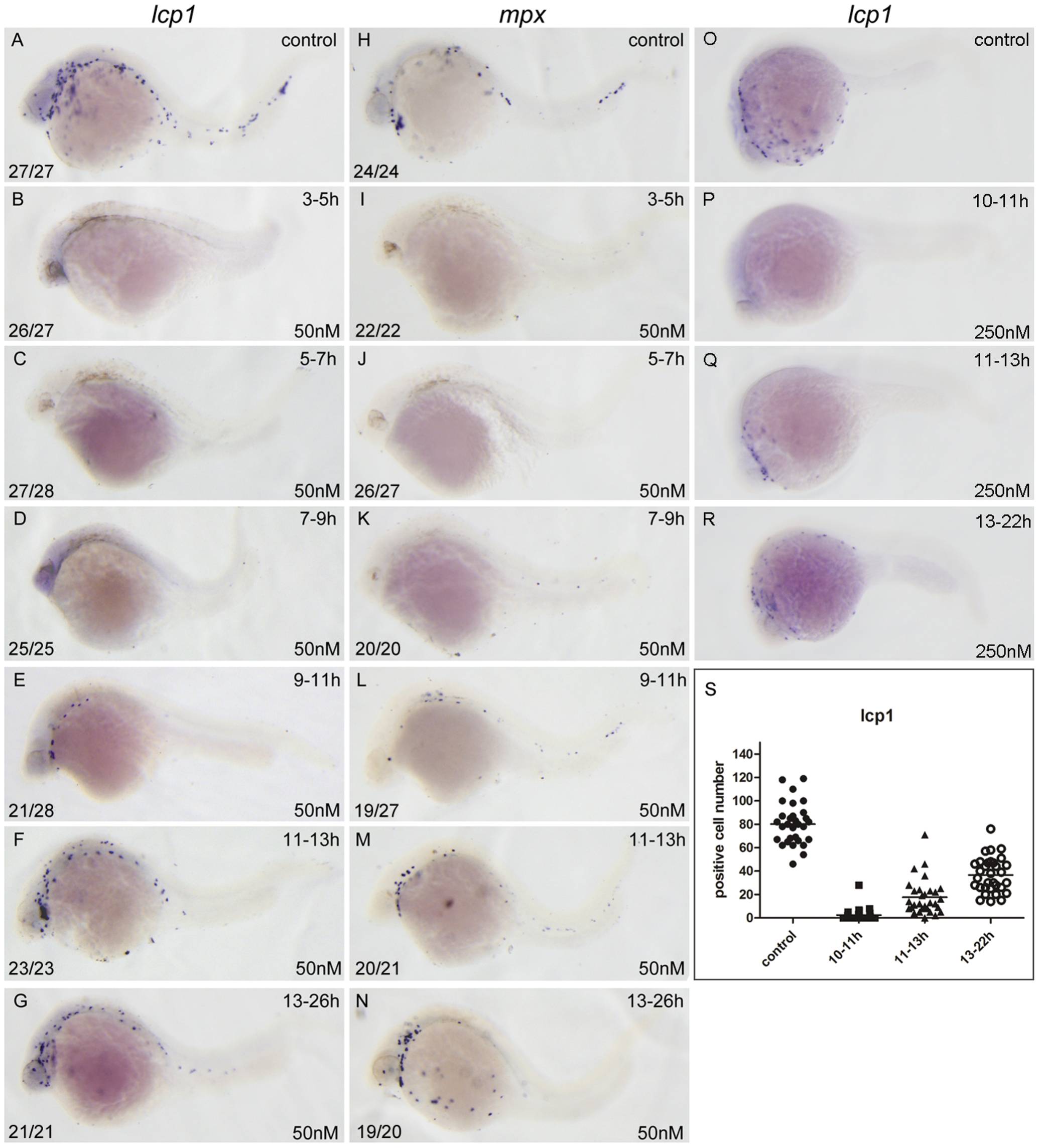
Fig. 2 RA restricts the primitive myelopoiesis mainly before 11 hpf.
All embryos are positioned anterior left and lateral front. Embryos were treated with vehicle DMSO (A, H, O) or with 50 nM RA (B–G, I–N) from 3 to 5 (B, I), 5 to 7 (C, J), 7 to 9 (D, K), 9 to 11 (E, L), 11 to 13 (F, M) and 13 to 26 hpf (G, N), or with 250 nM RA form 10 to 11 (P), 11 to 13 (Q), 13 to 22 hpf (R), respectively. They were then examined for expressions of myeloid markers lcp1 (A–G, O–R) and mpx (H–N) at 26 hpf (A–N) or 22 hpf (O–R) by whole mount in situ hybridization. The number shown in the lower left-hand corner of each panel is the number of embryos exhibiting the typical phenotype shown in the panel to the number of embryos totally observed. The typical embryos expressing lcp1+ cells at 22 hpf were shown in O–R. The scatter plot (S) shows the number of lcp1+ cells counted from each of the embryos at 22 hpf with different treatment (control; 10–11 hpf RA treatment; 11–13 hpf RA treatment and 13–22 hpf RA treatment).