
|
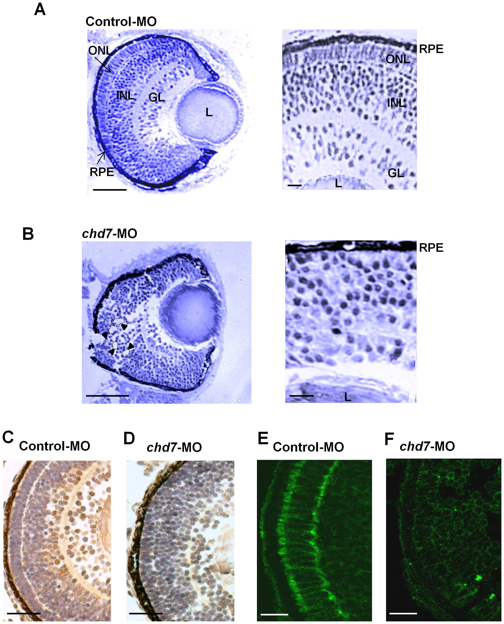
Fig. 7 Chd7 plays an essential role in retinal development.
Retinal organization of control-MO embryos (A) and chd7 morphants (B) was revealed by toludiene blue staining. Compared with the highly organized cells and laminated retinal structure in control-MO fish, chd7-MO retinal cells are disorganized (A–B: Left panels). Retinal lamination defect, including rosette formation,is clearly visible in the chd7 morphants (examples of rosettes are indicated by dotted lines and arrow heads). GL, ganglion cell layer; INL, inner nuclear layer; L, lens; ONL, outer nuclear layer; R, retina; RPE, retinal pigment epithelium. Scale bar: 50 μm. Zn-8 immunoreactivity was performed to label (brown) retinal gangion cells in control-MO (C) and chd7-MO embryos (D). Scale bar: 30 μm. The expression of retinal ganglion cell-specific marker zn-8 is greatly reduced in chd7 morphants. The photoreceptor layer of control-MO (E) and chd7-MO-injected (F) embryos were stained with 3A10. Chd7 morphants lacked the photoreceptor layer. Scale bar: 5 μm.