
|
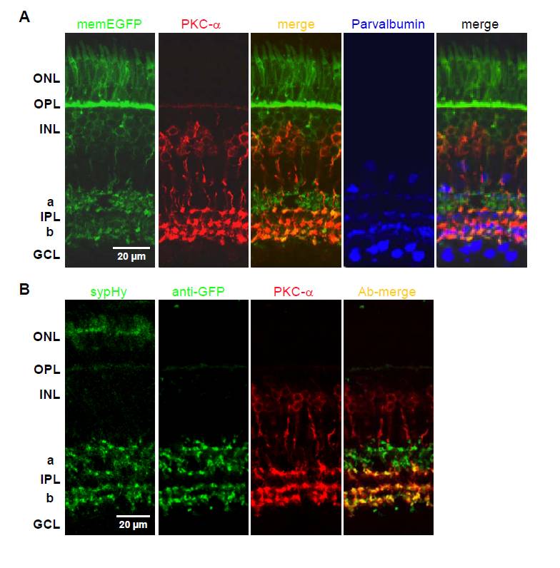
Fig. S1 Expression of fluorescent proteins in the zebrafish retina under the ribeye a (ctbp2) promoter
(A) Confocal images of a retinal section from a stable transgenic line of fish (Tg(- 1.8ctbp2:memEGFP)lmb) expressing membrane-targetted EGFP under the ribeye a promoter (8 dpf). The image shows a densely packed layer of photoreceptors in the outer nuclear layer (ONL), their terminals in the outer plexiform layer (OPL), cell bodies of bipolar cells in the inner nuclear layer (INL) and their terminals in the inner plexiform layer (IPL, sublamina a and b). Note the complete overlap between memEGFP and ON bipolar cells positive for PKCα staining (red). The neurons stained blue are positive for parvalbumin, a marker of A2 amacrine cells and displaced amacrine cells in the ganglion cell layer (GCL): note that the ribeye a promoter does not drive expression in these (merged image to right). (B) Confocal images of a retinal section from a stable transgenic line of fish (Tg(- 1.8ctbp2:sypHy)lmb) expressing sypHy under the ribeye a promoter (10 dpf). SypHy expression has been marked with an anti-GFP antibody. Expression is driven in photoreceptor terminals in the outer plexiform layer (OPL), and in bipolar cell terminals in the inner plexiform layer (IPL, sublamina a and b). There was complete overlap of sypHy expression and anti-PKCα staining (red, ON-bipolar terminals) in sublamina b of the IPL, while PKCα negative terminals were mainly situated in sublamina a (OFF-bipolar terminals). There is also green auto-fluorescence in photoreceptor outer segments.