
Image
|
Figure Caption
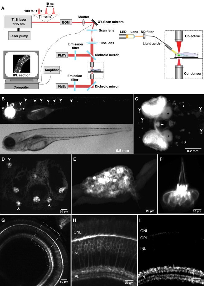
Fig. 1 The Zebrafish ribeye a (ctbp2) Promoter Drives Expression in Neurons Containing Ribbon Synapses
(A) Imaging synaptic reporters in the retina of live zebrafish using a two-photon microscope. Full-field stimuli were applied through a light guide.(B and C) A stable transgenic fish expressing membrane targeted EGFP (memEGFP) under control of 1.8 kb of the genomic sequence upstream of the ribeye a gene (Tg(-1.8ctbp2:memEGFP)lmb). At 4 dpf, all sensory organs known to express ribbon synapses were labeled, including the retina, the inner ear (white asterisk), the pineal gland (bold arrow), and the neuromasts (arrow heads). (B) Side view, (C) top view of the fish head. Additionally EGFP expression can be seen in the optic nerve and the optic tectum (black asterisk).(D) In a fish at 7 dpf, EGFP expression is driven in hair cells of the inner ear (side view) and maculae (not shown).(E and F) EGFP expression in the pineal gland and a neuromast, respectively (side view; 7 dpf).(G) In the retina, the ribeye a promoter drove expression of memEGFP in photoreceptors and bipolar cells.(H) Labeled photoreceptors in the outer nuclear layer (ONL), their terminals in the outer plexiform layer (OPL), cell bodies of bipolar cells in the inner nuclear layer (INL), and their terminals in the inner plexiform layer (IPL).(I) Expression of sypHy localized to terminals in the OPL and IPL in the stable Tg(-1.8ctbp2:sypHy)lmb line used in this study (see also Figure S1).
Acknowledgments
This image is the copyrighted work of the attributed author or publisher, and
ZFIN has permission only to display this image to its users.
Additional permissions should be obtained from the applicable author or publisher of the image.
Full text @ Neuron