
|
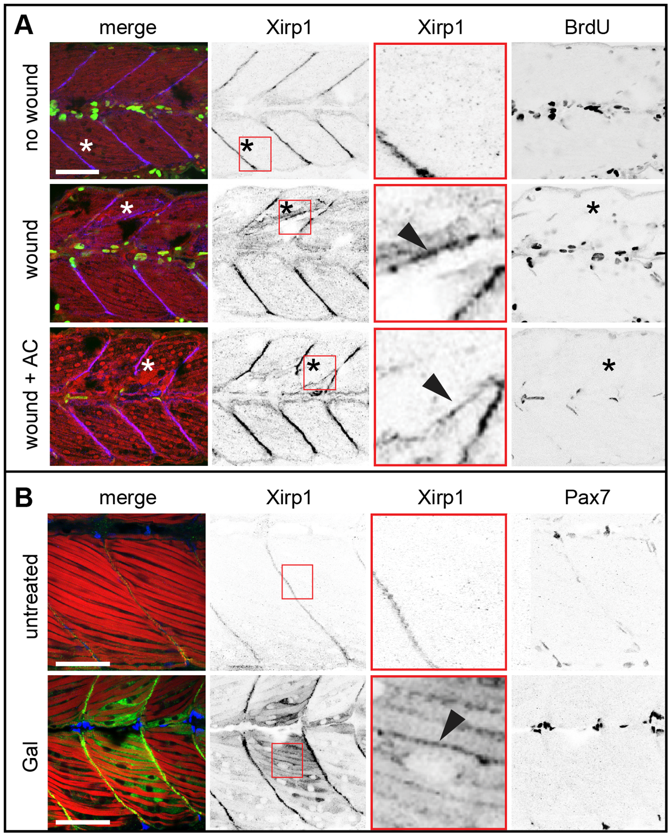
Fig. 4
Xirp1 marks wounded skeletal muscle cells prior to de novo cell proliferation.
(A) Laser-induced myocellular injuries in 33 hpf old zebrafish embryos after BrdU pulse labeling (between 24–33 hpf). Embryos were wounded at 31 hpf and left to recover for 2 hours. Xirp1+ tissue is devoid of BrdU+ proliferative cells. Treatment of laser-induced myocellular injuries with the proliferation inhibitor Aphidicolin (together with BrdU between 24–33 hpf) does not affect expression and localization of Xirp1 within damaged tissue. The efficacy of the Aphidicolin treatment is evident from strongly reduced BrdU labelling. Asterisks indicate the position of laser-induced injury within damaged tissue. Red inserts show Xirp1 localization within damaged tissue. Green: BrDU; red: Actin; blue: Xirp1. (B) Consistent with the lack of proliferating cells within Xirp1+ damaged tissue, the distribution of Pax7+ external cells is not changed compared to control conditions. Arrowheads mark ectopic Xirp1. Red inserts show Xirp1 localization within damaged tissue. Green: Xirp1; red: Actin; blue: Pax7. All scale bars: 50 μm.