
|
Fig. S5
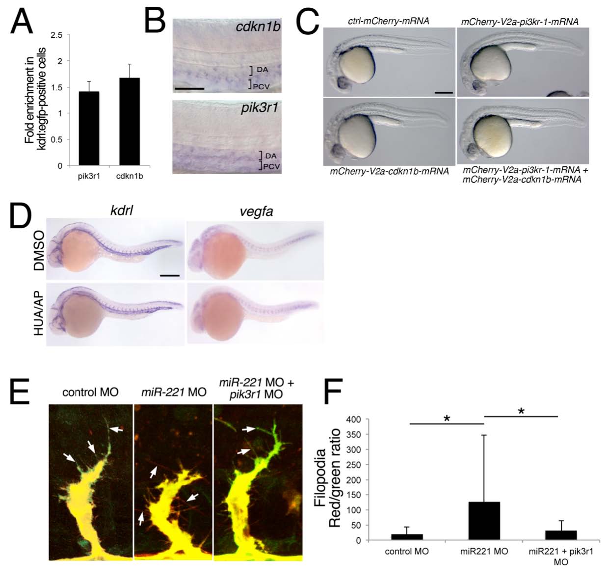
Proliferation and PI3 kinase signaling is required for ISV growth.
(A) Quantitative RT-PCR showing relative levels of expression of pik3r1 and cdkn1b in GFP+ versus GFPcells isolated from Tg(kdrl:egfp)la116 embryos at 24 hpf.
(B) Whole mount in situ hybridization detection of cdkn1b or pik3r1 in the dorsal aorta (DA) and posterior cardinal vein (PCV) at 24 hpf.
(C) Transmitted light images of embryos at 27 hpf injected with 500 pg mRNA encoding MCherry (control), Mcherry-2A-Cdkn1b, Mcherry-2A-Pikr1 or co-injected with 200 pg of mcherry-2A-cdkn1b and 300 pg of mcherry-2A-pik3r1 mRNA. Lateral views, dorsal is up, anterior to the left. Scale bar is 250 μm.
(D) Whole mount in situ hybridization of vegfa and kdrl in embryos treated with 4% DMSO, 150 μM 5- hydroxyurea (HUA) and 20 mM aphidocolin (AP), or 25 μM LY294002 beginning at 20 hpf. Scale bar is 250 μm.
(E) 2-photon microscopy of single ISVs in Tg(fli1ep:phaktegfp-2A-memcherry)um63;(kdrl:tagrfpcaax)is19 embryos injected with 15 ng control MO (left), 10 ng miR-221 MO (middle) or 5 ng pik3r1 MO and 10 ng miR- 221 MO (right). Filopodia are denoted by arrows. Embryos at 24 hpf (control MO) or 28 hpf (miR-221 and miR-221/pik3r1 MO). Scale bar is 10 μm.
(F) Quantification of red and green fluorescence in ISV filopodia following injection with indicated Morpholinos as in (E). Values indicate relative ratio of red (control) over PH-Akt1-EGFP fluorescence. Fluorescence was measured in at least 20 filopodia from 3 ISVs each in 3 embryos for each manipulation. An increased ratio is indicative of decreased PH-Akt1-EGFP localization in filopodia. * p< 0.05.
Reprinted from Developmental Cell, 22(2), Nicoli, S., Knyphausen, C.P., Zhu, L.J., Lakshmanan, A., and Lawson, N.D., miR-221 Is Required for Endothelial Tip Cell Behaviors during Vascular Development, 418-429, Copyright (2012) with permission from Elsevier. Full text @ Dev. Cell