
|
Fig. 8
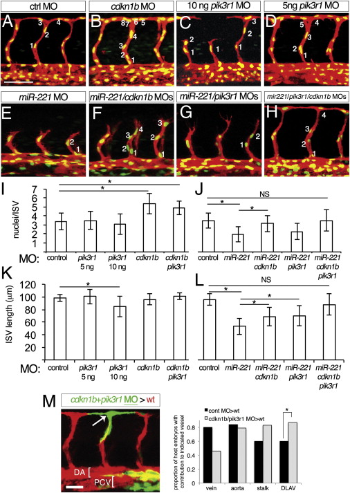
pik3r1 and cdkn1b Are Functional Downstream Targets of miR-221 (A–H) Tg(fli1a:negfp)y7;(kdrl:ras-cherry)s916 embryos at 27 hpf. Numbers denote cell nuclei of representative ISV. All embryos were co-injected with 1 ng of p53 MO. Scale bar is 50 μm. Embryo injected with (A) 15 ng control MO (B) 5 ng cdkn1b MO, (C) 10 ng pik3r1 MO, (D) 5 ng pik3r1 MO, (E) 10 ng miR-221 MO, (F) 10 ng miR-221 and 5 ng cdkn1b MOs, (G) 10 ng miR-221 and 5 ng pik3r1 MOs, and (H) 10 ng miR-221, 3 ng cdkn1b, and 3 ng pik3r1 MOs. (I–L) (I and J) Quantification of cells per ISV and (K, L) ISV length in embryos at 27 hpf injected with MOs as above, except control MO, which was injected at (I and K) 10 ng and (J and L) 15 ng doses. -p < 0.001; N.S., not significant. Measurements were obtained from four adjacent ISVs in at least ten embryos from three separate experiments. (M) Left, host Tg(kdrl:ras-mcherry)s916 embryo at 27 hpf following transplantation of cells from a Tg(fli1a:egfp)y1 embryo injected with 3 ng cdkn1b MO and 5 ng pik3r1 MO. Right, proportion of embryos displaying donor cells in indicated vessel type. Data from control donor cells is the same as in Figure 4D. -p = 0.07. Scale bar is 25 μm.
Reprinted from Developmental Cell, 22(2), Nicoli, S., Knyphausen, C.P., Zhu, L.J., Lakshmanan, A., and Lawson, N.D., miR-221 Is Required for Endothelial Tip Cell Behaviors during Vascular Development, 418-429, Copyright (2012) with permission from Elsevier. Full text @ Dev. Cell