
|
Fig. 6
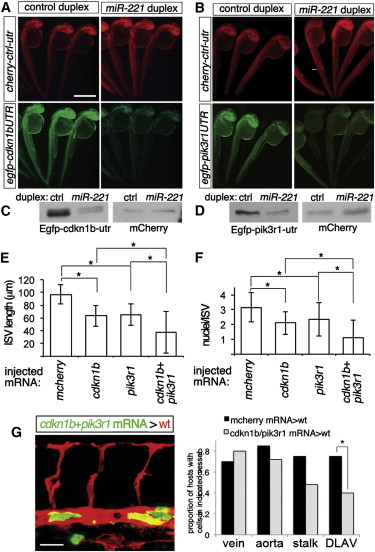
cdkn1b and pik3r1 Are Targets of miR-221 (A and B) Fluorescent images of embryos co-injected with 150 pg each of mcherry mRNA fused to control 32 UTR and egfp mRNA fused to (A) cdkn1b 32 UTR or (B) pik3r1 32 UTR with 100 μM indicated duplex RNA. Lateral views, dorsal is up, anterior to the left. Scale bar is 500 μm. (C and D) Western analysis of embryos in (A) and (B). (E and F) (E) ISV length and (F) cell number in Tg(fli1:egfp)y1 and Tg(fli1:negfp)y7 embryos, respectively, at 27 hpf injected with 500 pg of indicated mRNAs. For coinjections, 200 pg of cherry2A-cdkn1b and 300 pg cherry2A-pik3r1 mRNA were used. -p < 0.002. (G) Left, host Tg(kdrl:ras-mcherry)s916 embryo at 27 hpf following transplantation of cells from a Tg(fli1a:egfp)y1 embryo injected with 200 pg cdkn1b and 300 pg pik3r1 mRNA Right, proportion of embryos displaying donor cells in indicated vessel type. Control donor cells were from embryos injected with 500 pg mcherry mRNA. -p = 0.03. Scale bar is 25 μm
Reprinted from Developmental Cell, 22(2), Nicoli, S., Knyphausen, C.P., Zhu, L.J., Lakshmanan, A., and Lawson, N.D., miR-221 Is Required for Endothelial Tip Cell Behaviors during Vascular Development, 418-429, Copyright (2012) with permission from Elsevier. Full text @ Dev. Cell