
|
Fig. 5
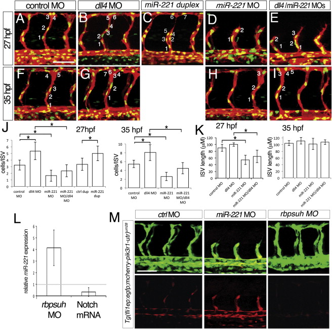
miR-221 Is Required for the Notch-Deficient “Hyper”-Angiogenesis Phenotype (A–I), Confocal micrographs of Tg(fli1a:negfp)y7;(kdrl:ras-cherry)s916 embryos, lateral views, dorsal is up, anterior to the left. Numbers indicate nuclei in representative intersegmental blood vessels (ISV). (A–E) Embryos at 27 hr post fertilization (hpf). (F–I) Embryos at 35 hpf. (A and F) Embryos injected with 25 ng of control MO. (B and G) Embryos injected with 15 ng of dll4 MO. (C) Embryo injected with 500 μM miR-221 duplex. (D and H) Embryo injected with 10 ng of miR-221 MO. (E and I) Embryos coinjected with 15 ng of dll4 MO and 10 ng of miR-221 MO. (J and K) (J) Quantification of endothelial number and (K) ISV length in Tg(fli1:ngfp)y7;(kdrl:ras-cherry)s916 embryos injected with indicated MO(s); -p < 0.0001. Measurements were determined for four adjacent ISVs per embryo in at least eight embryos from three separate injections. (L) Fold change of mature miR-221 levels assessed by qRT-PCR in embryos injected with 2.5 ng rbpsuh MO or with mRNA encoding an activated form of Notch compared to control embryos. (M) Confocal micrographs of Tg(fli1ep;mcherry-pik3r1-utr)um28 embryos injected with 10 ng control MO, 10 ng miR-221 MO, or 2.5 ng Rbpsuh MO. Lateral views, dorsal is up, anterior to the left. Scale bars are 50 μm.
Reprinted from Developmental Cell, 22(2), Nicoli, S., Knyphausen, C.P., Zhu, L.J., Lakshmanan, A., and Lawson, N.D., miR-221 Is Required for Endothelial Tip Cell Behaviors during Vascular Development, 418-429, Copyright (2012) with permission from Elsevier. Full text @ Dev. Cell