
IMAGE
Fig. S1
- ID
- ZDB-IMAGE-120216-71
- Genes
- Publication
- Yoruk et al., 2012 - Ccm3 functions in a manner distinct from Ccm1 and Ccm2 in a zebrafish model of CCM vascular disease
- All Figures
- Figures for Yoruk et al., 2012
Image
|
Figure Caption
Fig. S1
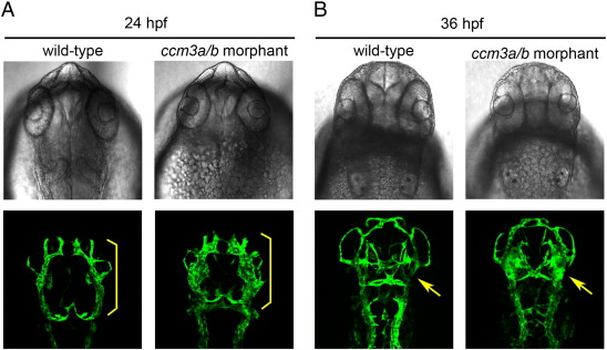
Cranial vasculature defects can be observed in ccm3a/b morphants at 24 and 36 hpf. At 24hpf, ccm3a/b morphants (0.75 ng ccm3a/b MO) display enlarged primordial midbrain channels (PMCs), compared to wildtype embryos (yellow brackets). At 36hpf, morphant midcerebral veins, which sprout from the PMC, also appeared enlarged compared to wildtype (yellow arrows).
Figure Data
Acknowledgments
This image is the copyrighted work of the attributed author or publisher, and
ZFIN has permission only to display this image to its users.
Additional permissions should be obtained from the applicable author or publisher of the image.
Reprinted from Developmental Biology, 362(2), Yoruk, B., Gillers, B.S., Chi, N.C., and Scott, I.C., Ccm3 functions in a manner distinct from Ccm1 and Ccm2 in a zebrafish model of CCM vascular disease, 121-131, Copyright (2012) with permission from Elsevier. Full text @ Dev. Biol.