
|
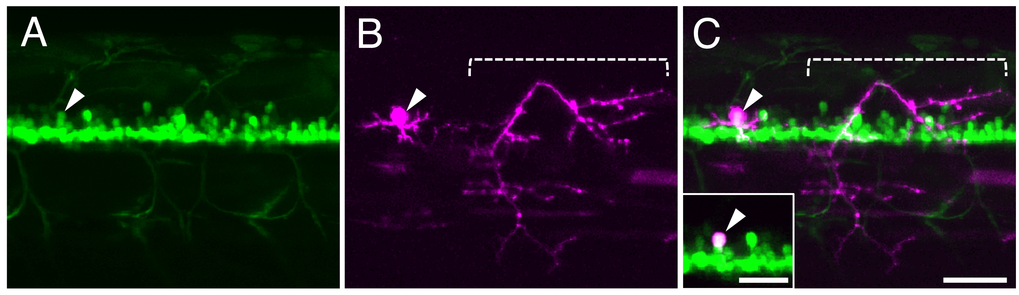
Fig. S1
Single-cell labeling of the spinal motor neuron in HGj4A larvae. A–C: The lateral view of the trunk region of the HGj4A larvae injected with the mnx1/hlxb9:mCherry plasmid. The arrowhead indicates the soma of a GFP-positive cell expressing RFP that locates slightly dorsal to the densely aligned GFP-positive cells in the ventral spinal cord. The dashed bracket indicates the motor axon innervating the skeletal muscles. In C, the GFP signal (A) and was merged into the RFP (B) signal. The inset in C shows the image of the same larvae taken with a shorter exposure time for RFP and indicates the single GFP-positive cell (arrowhead) is expressing RFP. Scale bars = 50 μm in A–C, 30 μm in the inset.