
Fig. 5
- ID
- ZDB-IMAGE-120216-32
- Genes
- Publication
- Balczerski et al., 2012 - Analysis of Sphingosine-1-phosphate signaling mutants reveals endodermal requirements for the growth but not dorsoventral patterning of jaw skeletal precursors
- All Figures
- Figures for Balczerski et al., 2012
|
Fig. 5
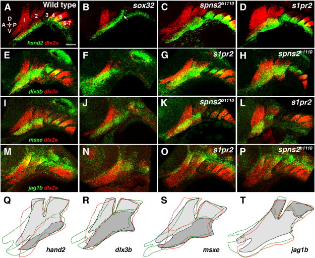
DV gene expression in endoderm mutants. (A–P) Double fluorescent in situ hybridizations show the expression of dlx2a (red) with hand2, dlx3b, msxe, or jag1b (green) in the pharyngeal arches (numbered) at 36 hpf. In wild types, hand2 (A) was restricted to the ventral-most domain, dlx3b (E) and msxe (I) to a DV-intermediate domain, and jag1b (M) to the dorsal arches. hand2 expression remained restricted to ventral arch CNCCs in sox32 embryos, although ectopic hand2 expression was seen in ectoderm overlying the second pouch (arrow) (B, n = 12/12). The hand2 expression domain remained ventrally restricted but was expanded in size in s1pr2 (D, n = 10/10), and spns2b1110 (C, n = 10/10) embryos. The DV-intermediate expression of dlx3b was normal in sox32 (F, n = 4/4), s1pr2 (G, n = 10/10), and spns2b1110 (H, n = 5/5) embryos, as was expression of msxe in sox32 (J, n = 7/7), s1pr2 (L, n = 11/11), and spns2b1110 (K, n = 5/5) embryos. jag1b expression was reduced but remained dorsal-restricted in sox32 embryos (N, n = 5/5) and was unaffected in s1pr2 (O, n = 5/5) and spns2b1110 (P, n = 6/6) embryos. (Q–T) Tracings of the dlx2a-expressing arches (solid lines) and the DV expression borders (dashed lines) of hand2 (Q), dlx3b (R), msxe (S), or jag1b (T) show slight expansion of hand2 expression but no change in dlx3b, msxe, or jag1b expression in two classes of s1pr2 and spns2b1110 mutants. For wild types, dlx2a expression is light gray, with DV gene expression domains in darker gray. The mandibular arch was expanded (green lines) in some embryos (C, G, K, O; 47% of s1pr2 and 53% of spns2b1110) and was abnormally shaped and/or anteriorly truncated (red lines) in other embryos (D, H, L, P; 47% of s1pr2 and 16% of spns2b1110). Scale bar = 50 μm.
Reprinted from Developmental Biology, 361(2), Balczerski, B., Matsutani, M., Castillo, P., Osborne, N., Stainier, D.Y., and Crump, J.G., Analysis of Sphingosine-1-phosphate signaling mutants reveals endodermal requirements for the growth but not dorsoventral patterning of jaw skeletal precursors, 230-241, Copyright (2012) with permission from Elsevier. Full text @ Dev. Biol.