
|
Fig. 2
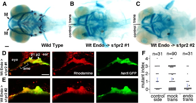
Wild-type endoderm rescues lower jaw development in s1pr2 mutants. (A–C) Ventral views of 6 dpf head skeletons. Unilateral transplantation of wild-type endoderm precursors into s1pr2 mutants rescued the loss (B) or reduction (C) of lower jaw M cartilage seen in control sides not receiving transplants. (D and E) The same two examples imaged at 30 hpf to show the contribution of wild-type her5:GFP+ (green) rhodamine-dextran+ (red) donor endoderm cells to the anterior medial endoderm (ame) and the first and second pouches (p1 and p2) of the host arches. (F) For quantification of lower jaw rescue, we devised a mutant index: 0, wild type; 1, minor M reduction; 2, major M reduction; 3, M missing; 4, M and Pq missing. Compared to s1pr2 mutants receiving mock transplants, mutant sides receiving endoderm transplants (but not the contralateral control sides of the same animals) had partial restoration of lower jaw cartilage. Individual data points are plotted, and bars show standard error of the mean. Asterisks denote the two groups with statistical differences in average jaw skeletal defects. Scale bars = 50 μm.
Reprinted from Developmental Biology, 361(2), Balczerski, B., Matsutani, M., Castillo, P., Osborne, N., Stainier, D.Y., and Crump, J.G., Analysis of Sphingosine-1-phosphate signaling mutants reveals endodermal requirements for the growth but not dorsoventral patterning of jaw skeletal precursors, 230-241, Copyright (2012) with permission from Elsevier. Full text @ Dev. Biol.