
|
Fig. S5
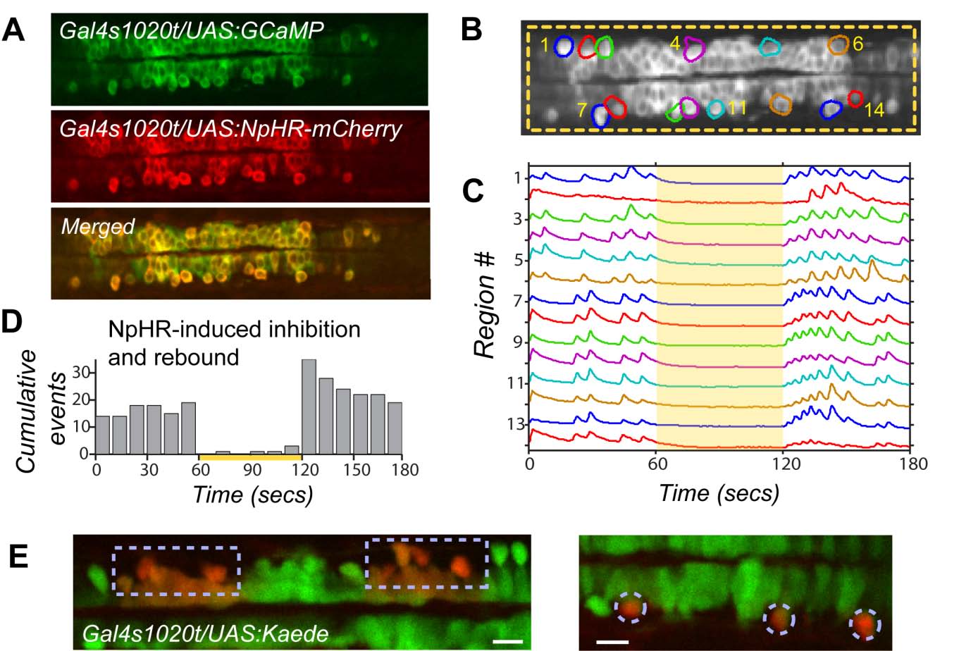
Activation of the Light-Driven Chloride Pump Halorhodopsin Robustly Inhibits Spontaneous Events and Causes Rebound Excitation, Related to Figures 4 and 5
(A) Gal4s1020t/ UAS:GCaMP3/ UAS:NpHR-mCherry fish express GCaMP3 and NpHR in the same spinal neurons.
(B) Full field illumination (yellow dashed box) with 19mW/mm2 593nm light is targeted to NpHR-expressing spinal neurons while simultaneously imaging population activity with GCaMP3.
(C) Spontaneous events in active regions, plotted as standard deviation, are eliminated during application of yellow light (yellow bar) in a 19 hpf fish.
(D) Tallied events across several fish (n=6) shows effective inhibition when yellow light is on (yellow bar) and there is an increase in activity above baseline at light offset.
(E) Accurate light targeting with the digital micro-mirror device is confirmed with the restricted photo-conversion of Kaede. Illumination with blue light (380nm) switches the fluorescence emission of Kaede from green to red22. Cells within illuminated region (boxed areas on left, single cells circled on right) convert to red while adjacent cells remain green. Though the wavelength used for NpHR activation is longer (593nm) and may scatter a little differently, we used a similar light power (15.2mW/mm2 for Kaede; 19mW/mm2 for NpHR) and exposure time (1 min). Scale bars=20μm.