
Fig. 1
- ID
- ZDB-IMAGE-120210-2
- Publication
- van Eeden et al., 1996 - Genetic analysis of fin formation in the zebrafish, Danio rerio
- All Figures
- Figures for van Eeden et al., 1996
|
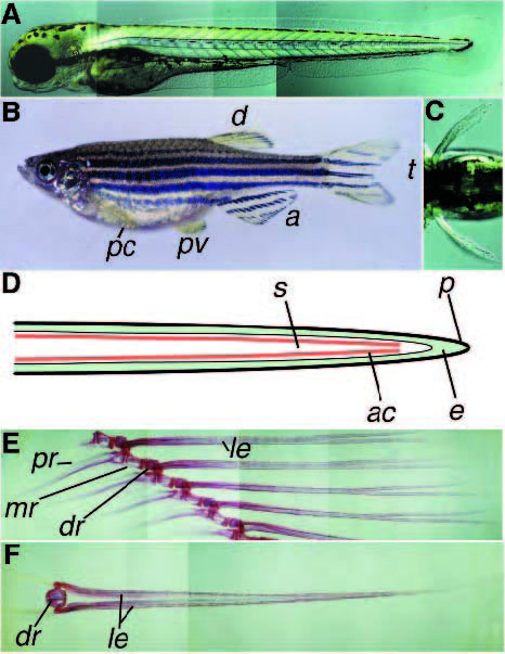
Fig. 1
Overview of wild-type fins in zebrafish. (A) Wild-type embryo at 84 hpf, caudal fin. (B) Adult female showing the five different types of fins. Going from anterior to posterior, the pectoral fins (pc) and the pelvic fins (pv), the dorsal fin (d), the anal fin (a) and the tail fin (t). (C) Dorsal view of an approximately 84 hpf wildtype embryo showing the pectoral fins. (D) Schematic drawing of a frontal section through an embryonic fin. Epiderm (e, green) is covered by periderm (p, black), in the subepidermal space (s) between the two epidermal layers actinotrichia (ac, red) are present in a double layer. Actinotrichia are also visible in Fig. 2A. (E) Skeletal staining of a part of the adult anal fin and the supporting skeleton. The endoskeletal radials consist of the proximal (pr), the medial (mr) and a distal (dr) part. The segmented lepidotrichia (le), belonging to the dermal skeleton are connected to the distal radial. (F) Frontal view of one adult anal fin segment showing the bilaterally arranged lepidotrichia connected to the distal radial (dr).