
|
Fig. S1
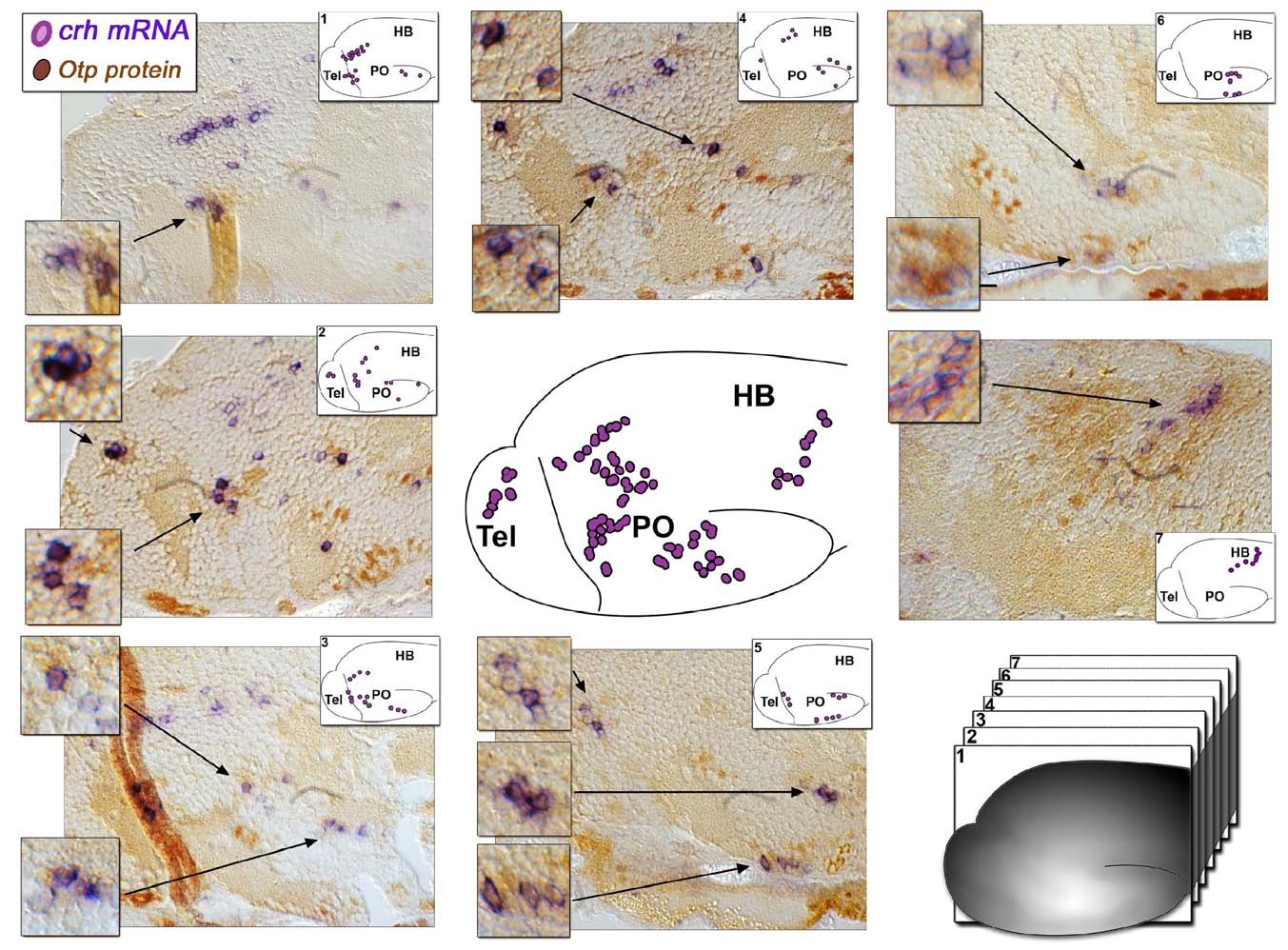
Expression of Otp and CRH in 6-day-old Zebrafish Larva, Related to Figure 1
Serial sagittal sections (6 μm) of 6-day old zebrafish larva subjected to whole mount in situ hybridization with a crh- directed probe (purple) followed by paraffin embedment and sectioning, antigen retrival and immuno-staining with an anti-Otp antibody (brown). The scheme in the center of the figure depicts the location of the major CRH+ neuronal clusters in 6-day old larva, whereas schemes shown in the insets of the image panels depict the subsets of CRH+ cells in each of the tissue sections. High-magnifications of crh+;Otp+ neuronal clusters (arrows) are also presented.