
Fig. 3
- ID
- ZDB-IMAGE-120202-9
- Publication
- Kwan et al., 2012 - A complex choreography of cell movements shapes the vertebrate eye
- All Figures
- Figures for Kwan et al., 2012
|
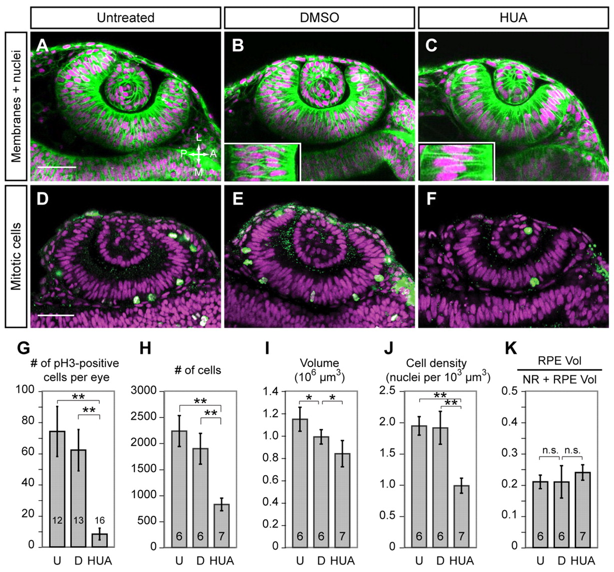
Fig. 3
Proliferation is dispensable for OCM. (A-C) Confocal sections (live, 24-hpf) showing EGFP-CAAX (membranes, green) and H2A.F/Z-mCherry (nuclei, magenta). (A) Untreated, (B) DMSO and (C) HUA embryos treated from 10.5-24 hpf. Insets show large HUA-treated cells versus DMSO control. (D-F) Confocal sections at 24 hpf showing phospho-histone H3 (green) and TO-PRO-3 (magenta) in (D) untreated, (E) DMSO-treated and (F) HUA-treated embryos. Dorsal view. (G-K) Quantitative analyses of (G) mitoses, (H) cell number, (I) OV volume, (J) cell density and (K) retinal pigmented epithelium (RPE) volume as a fraction of OC [neural retina (NR) plus RPE] at 24 hpf in untreated (U), DMSO-treated (D) and HUA-treated embryos. Numbers of embryos scored (one eye each) are indicated within each bar. *, P<0.05; **, P<0.01; error bars indicate s.d. Scale bars: 50 μm.