
Image
|
Figure Caption
Fig. S1
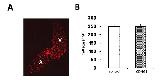
related to Figure 3: Cardiac cell size is not altered in CDNG1-treated embryos. (A) Fluorescent optics exhibiting individual nuclei of cardiomycoytes in flat-mounted Tg(cmlc2:DsRed-nuc) embryos (60hpf). (B) Cardiac cell size is comparable in embryos treated by CDNG1 at 30μM (5-60 hpf) compared to control embryos. 20 cardiomyocytes were chosen from 3 treated and untreated embryos for cell size analysis. Graph shows mean ±s.d.
Acknowledgments
This image is the copyrighted work of the attributed author or publisher, and
ZFIN has permission only to display this image to its users.
Additional permissions should be obtained from the applicable author or publisher of the image.
Full text @ Chem. Biol.