
|
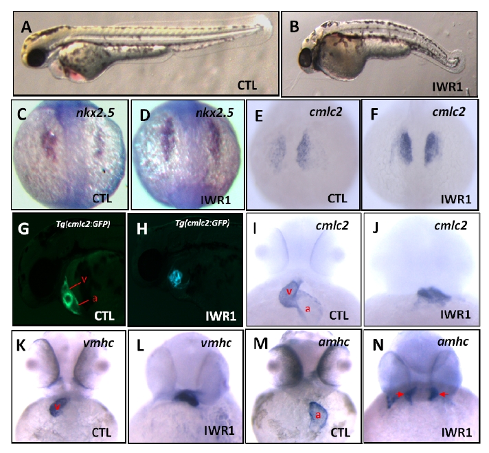
Fig. S3
related to Figure 6: IWR1 treatment causes defects in zebrafish embryo and heart development. (A) Lateral view displaying a wild-type embryo with a normal body length. (B) Embryos treated with IWR1 exhibiting tail truncation in loosing tail structure posterior to the yolk extension. (C, D) In situ hybridization analyses revealed normal nkx2.5 expression in a wild-type embryo and increased nkx2.5 expression in a IWR1-treated embryo. (E, F) Dorsal views displaying the normal cmlc2 expression in a wild-type embryo and increased cmlc2 expression in a IWR1-treated embryo. (G, H) Fluorescent optics in lateral views exhibiting a normal atrium and a normal ventricle in a wild-type Tg(cmlc2:EGFP) embryo and defective cardiac chambers in a IWR1-treated Tg(cmlc2:EGFP) embryo. (I, J) cmlc2 expression in ventral views revealing a normal atrium and ventricle in a wild-type embryo and defective cardiac chambers in a IWR1-treated embryo. (K, L) vmhc expression in ventral views showing a normal ventricle in a wild-type embryo and a defective ventricle in a IWR1-treated embryo. (M, N) amhc expression in ventral views exhibiting a normal atrium in a wild-type embryo, and bilateral-aligned atrial myocytes (red arrows) in a IWR1-treated embryo, leading to failure to form the atrium. a: atrium; v: ventricle; IWR1: 20 μM. 12 hpf (C, D); 17 hpf (E, F); 24 hpf (A, B); 48 hpf (G-N).