
Fig. 8
- ID
- ZDB-IMAGE-120202-14
- Publication
- Kwan et al., 2012 - A complex choreography of cell movements shapes the vertebrate eye
- All Figures
- Figures for Kwan et al., 2012
|
Fig. 8
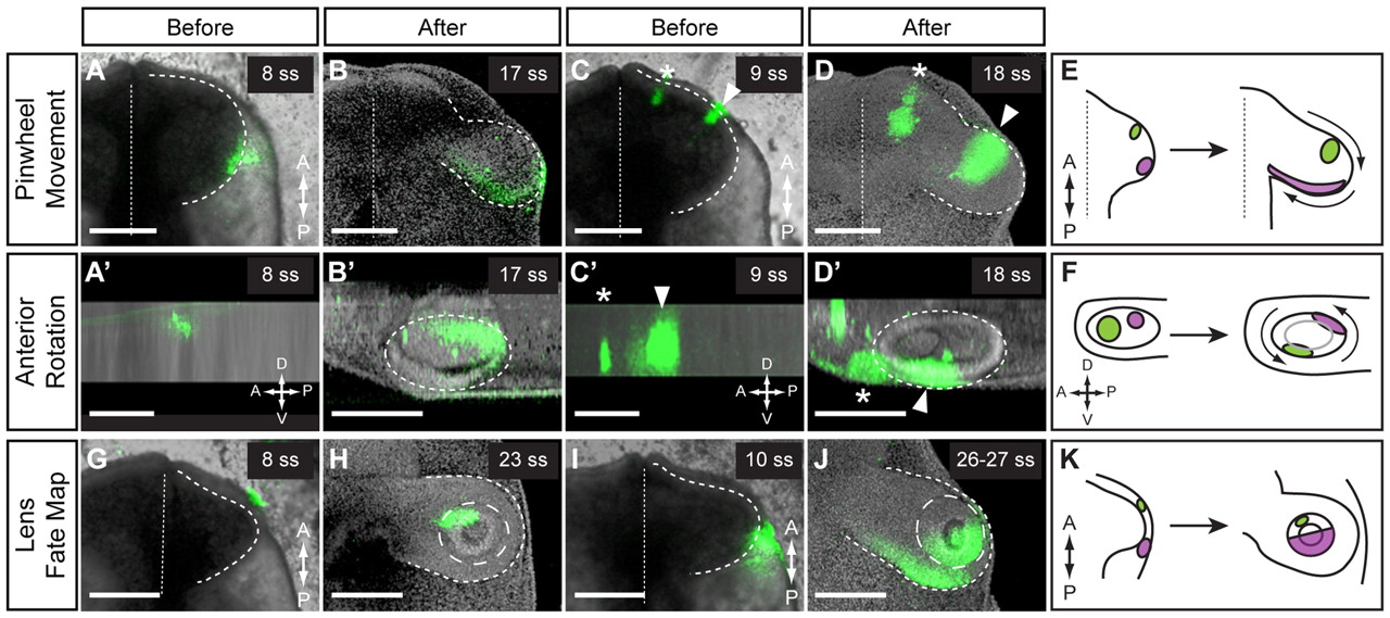
Conservation of movements in chick OCM. (A-F) Pinwheel movement and anterior rotation. Lateral (A,A2) or anterior (C,C2) OV was labeled and imaged immediately. (A,C) Ventral views. (A2,C2) Lateral views. (C) Two anterior DiI spots were marked: medial spot (asterisk), brain; lateral spot (arrowhead), OV. The lateral DiI spot moved medially (B) and dorsally (B2). The anterior DiI spot (arrowhead) moved laterally (D) and ventrally (D2). (B,D) Ventral views. (B2,D2) Lateral views. Dashed line outlines OV; dotted line indicates the midline. (E) Summary of pinwheel movement. (F) Summary of anterior rotation. (G-K) Lens fate map. Anterior (G) or posterior (I) ectoderm was labeled and imaged immediately. Ventral view. Dashed line outlines OV; dotted line indicates the midline. (H,J) The DiI label was found in distinct lens domains. Ventral view. Dashed line outlines OV; long-dashed line outlines lens. (K) Summary of lens fate map. Volume renderings: DiI and brightfield (A,A2,C,C2,G,I) or DiI and nuclear counterstain (B,B2,D,D2,H,J). Scale bars: 200 μm.