
|
Fig. S3
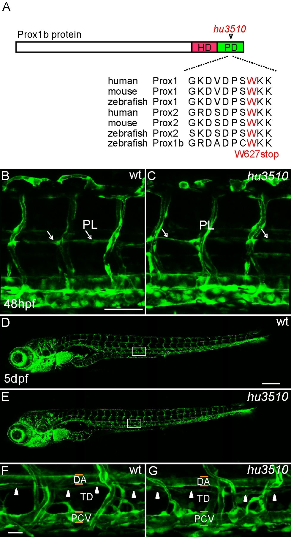
The lymphatic development of homozygous prox1bhu3510 mutant is normal. (A) Schematic representation of the Prox1b protein, with the position of the prox1bhu3510 allele indicated. The homeodomain region is shown in red, the Prospero domain in green. The predicted stop mutation occurs at W627 in prox1bhu3510 and multiple sequence alignment shows the conservation of zebrafish W627 in the Prospero domain of Prox proteins. (B) and (C) show the vascular structures in the trunk region of wt (B) and homozygous prox1bhu3510 mutant embryos (C) in fli1:GFP background. The white arrows indicate PLs. (D) and (E) show the full images of 5-day wt (D) and homozygous prox1bhu3510 mutant embryos (E). (F) and (G) show enlarged views of the boxed areas in (D) and (E). The white arrowheads indicate the presence of TD in both control (F) and homozygous prox1bhu3510 embryos (G). Scale bars represent 50 μm in (B), 250 μm in (D) and 25 μm in (F).