
|
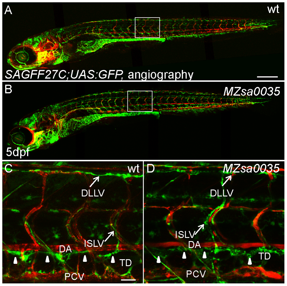
Fig. 5
The lymphatic development of maternal-zygotic prox1bsa0035 mutant is unaltered.
(A) and (B) show whole embryo lateral view images of 5-day wt (A) and MZ prox1bsa0035 mutant (B) in a SAGFF27C;UAS:GFP background. Perfused blood vessels were labeled by angiography (in red). (C) and (D) show enlarged views of the boxed areas in (A) and (B). The entire lymphatic network in the trunk of zebrafish, which is composed of the GFP-expressing lymphatic vessels-DLLV, ISLV and TD (marked by white arrowheads), is properly formed in wt (C) and MZ prox1bsa0035 mutant embryos (D). Scale bars represent 250 μm in (A), and 25 μm in (C).